Download

1 / 11
110 likes | 218 Vues
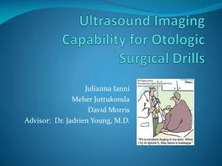
Ultrasound Imaging Capability for Otologic Surgical Drills. Julianna Ianni Meher Juttukonda David Morris Advisor: Dr. Jadrien Young, M.D. What is Otologic Surgery?. Surgery of the ear Mastoidectomy Mastoid air-filled spaces behind the ear Surgery to remove cells from the mastoid

E N D
Ultrasound Imaging Capability for Otologic Surgical Drills JuliannaIanni MeherJuttukonda David Morris Advisor: Dr. Jadrien Young, M.D.
What is Otologic Surgery? • Surgery of the ear • Mastoidectomy • Mastoid • air-filled spaces behind the ear • Surgery to remove cells from the mastoid • Uses • to treat anti-biotic resistant infections in the region • to insert a cochlear implant • 30,000 to 60,000 performed annually in the U.S.1
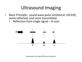
Objectives • To find an ultrasound transducer that is compatible with an otologic drill • To calculate the thickness of the mastoid bone using A-mode US • To shut off the drill when the bone has been drilled
Past Work • Studied ultrasound equipment in order to determine the most effective way to produce accurate images • Researched the best transducer frequency for imaging that region of the skull • Met with Dr. Young and discussed the surgical aspects required to have a usable drill • Read several papers and technical documentation regarding the operation of ultrasound surgical imaging technology • Developed the website • Updated list of design goals • Observed use of otologic drills & identify design constraints • Identified potential design obstacles • Generated design ideas concerning mechanism of attachment • Restructured design goals focusing more on finding an ultrasound transducer compatible with an otologic drill
Current work • Finding company that can build hollow annular transducer to specifications • Performing measurements w/larger transducer on material samples analogous to skull bone • Researching renting a ultrasound depth gauge to test on cadaver bone for proof of concept
Simulation of Signal • Assumptions • Speed of Sound in Skull Bone = 2700 m/s3 • Only Reflection/Transmission & Attenuation • No Scattering • Results
Solidworks Prototype Side View Top View Bottom View
Future Work • Deciding type & shape of ultrasound transducer • Determine ideal frequency • Developing method to connect to current power source • Developing cooling system • Developing a B-Mode to image the path and Doppler mode to measure blood flow • Building & testing prototype
References • 1. French, LC et al. “An estimate of the number of mastoidectomy procedures performed annually in the United States”. Ear Nose Throat J. 2008 May; 87(5): 267-70. • 2. Ear Anatomy: http://www.umm.edu/imagepages/1092.htm • 3. Clement, GT et. Al. “Correlation of Ultrasound Phase with Physical Skull Properties”. Ultrasound in Medicine & Biology. 2002 May; 28(5): 617-624.