Centromere
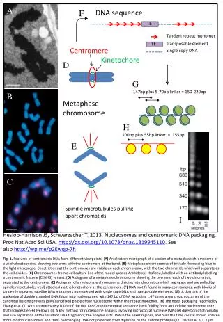
A. F. DNA sequence. TE. TE. Tandem repeat monomer. Transposable element. Single copy DNA. Centromere. Kinetochore. D. G. 147bp plus 5-70bp linker = 150-220bp. B. Metaphase chromosome. 100bp plus 55bp linker = 155bp. H. E. C. Spindle microtubules pulling apart chromatids. I.

Centromere
E N D
Presentation Transcript
A F DNA sequence TE TE Tandem repeat monomer Transposable element Single copy DNA Centromere Kinetochore D G 147bp plus 5-70bp linker = 150-220bp B Metaphase chromosome 100bp plus 55bp linker = 155bp H E C Spindle microtubules pulling apart chromatids I Heslop-Harrison JS, Schwarzacher T. 2013. Nucleosomes and centromeric DNA packaging. Proc Nat AcadSci USA. http://dx.doi.org/10.1073/pnas.1319945110. See also http://wp.me/p2Ewqp-7h Fig. 1. Features of centromeric DNA from different viewpoints. (A) An electron micrograph of a section of a metaphase chromosome of a wild wheat species, showing two arms with the centromere at the bend. (B) Metaphase chromosomes of triticale fluorescing blue in the light microscope. Constrictions at the centromeres are visible on each chromosome, with the two chromatids which will separate as the cell divides. (C) Chromosomes from a cell culture line of the model species Arabidopsis thaliana, labelled with an antibody labelling a centromerichistone (CENH3) variant. (D) A diagram of a metaphase chromosome showing the two arms each of two chromatids, separated at the centromere. (E) A diagram of a metaphase chromosome dividing into chromatids which segregate and are pulled by spindle microtubules (red) attached via the kinetochore at the centromere. (F) DNA motifs found in many centromeres, with blocks of tandemly repeated satellite DNA monomers interspersed with single copy DNA and transposable elements. (G). A diagram of the packaging of double stranded DNA (blue) into nucleosomes, with 147 bp of DNA wrapping 1.67 times around each octamer of the canonical histone proteins (olive) and fixed phase of the nucleosome within the repeat monomer. (H) The novel packaging reported by Zhang et al. (1) with approximately 100bp of the rice CentO tandem repeat sequence (red) folding once around the nucleosome core that includes CenH3 (yellow). (I). A key method for nucleosome analysis involving micrococcal nuclease (MNase) digestion of chromatin and size separation of the resultant DNA fragments; the enzyme cuts DNA in the linker regions, and over the time course shown isolates more mononucleosomes, and trims overhanging DNA not protected from digestion by the histone proteins (12). Bars in A, B, C 2 µm