Diatom Nanotechnology
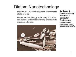
Diatom Nanotechnology. By Susan J. Crawford-Young Electrical & Computer Engineering, University of Manitoba, 2003, mudworks@access.cici.mb.ca. Diatoms are unicellular algae that form intricate shells of silica.

Diatom Nanotechnology
E N D
Presentation Transcript
Diatom Nanotechnology By Susan J. Crawford-Young Electrical & Computer Engineering, University of Manitoba, 2003, mudworks@access.cici.mb.ca Diatoms are unicellular algae that form intricate shells of silica. Diatom nanotechnology is the study of how to use diatoms or their silica forming processes to make nanodevices.
Diatoms are three dimensional. A scanning electron microscope is needed to see this. Frustule Frustules are made like two overlapping boxes. epitheca Septum hypotheca Diatoms range in size from 1 to 500 µm the ones in this picture are approx 20 µm
Diatoms come in many shapes and forms. There are approximately 105 species of diatom each with a different shape. 10µm
There are two types of diatoms Pinnate – Most species of this type of diatom can move. Centric The colors of the diatoms are partially due to the refraction of light in the silicon shells and partly due to their brown pigmentation. Diatoms are small refraction gratings and are good at collecting light.
Diatoms often collect in shaped colonies. This type of self assembly might be of use in building a nano device. A star made of pennatae diatoms Zipper Diatom Licmophora colony
Why Use Diatoms ? • Diatoms have nanometer features that are not reproducible by current technology • Diatoms grow themselves as long as there is light, heat and the correct nutrients available. • Grow at ambient temperatures and pressures. Problems with using Diatoms • Have to grow diatoms of one type or species to use in a specific application. • There is no automated way to place diatoms into a MEMS device. They have to be arranged by hand or persuaded to grow in the right place.
Growing the right Diatom Growth media with antibiotic Laser guided by a vision recognition system Genetically altered diatom antibiotic resistant Dead diatom Dead diatom Using Biotechnology Using a Compustat
Using diatoms as a template Kovacs, G.T.A.; “Micromachined Transducers Sourcebook” 1998 : page 137
Diatom Size Change Micrograph courtesy of M. B. Edlund Frustules of the diatom Stephandiscus niagarae Ehrenb., viewed in a scanning electron microscope. Larger frustule is complete, and is near maximum size range for this taxon. In the smaller frustule, which is near the minimum size range for the species, the valves are separated, and the interior of one valve is visible.
Diatom Growth Frustules divide (asexually reproduce) by separating and then each part produces another half. The bottom diatom will be slightly smaller. This causes a size range in diatoms. epitheca Septum hypotheca
Silica Starvation • - is a method of growing diatoms so that they are at the same stage. • Partially grown diatom shells could be made using this method. • The diatoms are grown in a Teflon container so that they do not have any silica available to use for growth. The diatoms then reproduce and grow only when silica is added. This is because DNA synthesis is silica dependent In diatoms. Teflon Diatoms in nutrients and light, without silica
Growing Diatoms on a Comb 1. 2. Make a silicone comb by etching Dip the comb is Teflon Diatoms 3. 4. Dip in silicon starved diatom mix. The diatoms will attach themselves to the silica via hydrophilic mucilage. Polish off the Teflon on the tips of the comb
Using silica starvation to grow a diatom comb Acknowledgement: Dr. R. Gordon Diatoms grow in several different ways. Pennate diatoms grow like this. Partly grown diatom New diatom shells grow inside the diatom cells in synchrony when silica is added to a silica starved diatom culture. If the diatoms are killed all at the same time, and at the right stage, there will be diatom combs in the mixture of shells. 80µm Partially grown diatoms are fragile so further processing might be necessary. A fully grown diatom like this starts off as a comb shape.
Diatom Diffraction Grating One way to create diffraction gratings using diatoms would be to align them, secure them in place, and then copy them with a molding technique. The molding technique has to be able to reproduce fine nanometer scale features. The ribs and diffraction gratings also form structural members.
Biosilicon • Biosilicon is the material that diatom shells are made of. • It is made of silicon silaffins, frustulins and polyamines. • It is made by precipitation SiO2 using polypeptides (silaffins, frustulins) • and polyamines. • comes in differing hardness in diatoms and therefore could be made • with varying hardness levels.
Structure of Silaffin 1A from C.fusiformis H2N—S—S—K—K—S—G—S—Y—S—G—S—K—G—S—K—COOH OH N N N+ N H H3C CH3 H3C CH3 H CH3 N N H n=4-9 H n=4-9 N N CH3 CH3 N N CH3 H3C CH3 H3C
Structure of Polyamines from N. angularis A family of 25 amines is generated by variation of the chain length and the degree of methylation. Additional variation is achieved by exchange of the butylamine (left) for a propylamine unit. R N R N N N n CH3 CH3 R R = CH3, H n = 7-11 Pohnert G., Angew Chem. Int. Ed. 2002, 41, No. 17
Silica precipitates induced by N angularis polyamides. Scale bars 500 nm (A and B) and 1 µm (C to F) • Polyamine molecular masses 1,000 to 1,200 Da used • (B) Polyamine molecular masses 600 to 750 Da used • (C - F) Polyamine molecular masses 600 to 1,250 Da used • (C) pH 5.4 • (D) pH 6.3 • (E) pH 7.2 • (F) pH 8.3
Effect on silica morphology of combining N. angularis polyamines and silaffins. • Enriched silaffins used for precipitation. • (B) Mixture of silaffins and polyamines used for precipitation. • Scale bars 500 nm and 1µm insets Kroger et al (1997) (26) 14133
Magnesium Diatom Shells Diatom shells were placed in an atmosphere of magnesium at 900o C for four hours. The overall frustule shape and meso/nanoscale features were retained. View of open end (other end closed) Other ceramic materials could be made using this technique. Research by Dr. Kenneth Sandhage Longitudinal view revealing fine ( 102 nm diameter Pores )
Radiolaria Another type of marine life form that has a silica shell. www.microscopy-uk.org.uk
Making microscopic art with diatoms. The End Reference web sites www.bgsu.edu www.calacademy.org hjs.geol.uib.no www.indiana.edu www.microscopy-uk.org.uk Canadian Museum of Nature