
Diagnosis Dilemma: Primary Apocrine Adenocarcinoma of the Scrotum vs. Urothelial Carcinoma
This case report discusses a rare instance of apocrine adenocarcinoma of the scrotum, initially suspected to be metastatic urothelial carcinoma. We present a 79-year-old male with a tender scrotal mass, following treatment for T1G3 urothelial carcinoma. Histological evaluation revealed a malignant tumor with papillary architecture. The diagnosis of apocrine adenocarcinoma was confirmed after considering alternative primary sites. The management included partial scrotectomy and selective lymphadenectomy, illustrating the challenges in diagnosis and treatment of such uncommon malignancies.

Diagnosis Dilemma: Primary Apocrine Adenocarcinoma of the Scrotum vs. Urothelial Carcinoma
E N D
Presentation Transcript
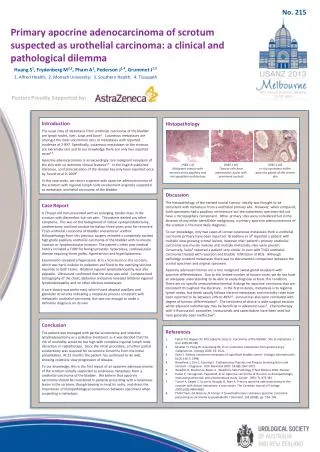
No. 215 Primary apocrine adenocarcinoma of scrotum suspected as urothelial carcinoma: a clinical and pathological dilemma Huang S1, Frydenberg M2,3, Pham A1, Pederson J1,4, Grummet J1,2 1. Alfred Health; 2. Monash University; 3. Southern Health; 4. Tissupath Posters Proudly Supported by: Introduction The usual sites of metastasis from urothelial carcinoma of the bladder are lymph nodes, liver, lungs and bone1. Cutaneous metastases are amongst the most uncommon sites of metastases with reported incidence of 2-8%2. Specifically, cutaneous metastases to the scrotum are extremely rare and to our knowledge there are only two reported cases3,4. Apocrine adenocarcinoma is an exceedingly rare malignant neoplasm of the skin with no definitive clinical features5,6. In the English published literature, scrotal localisation of the disease has only been reported once by Tuncel et al in 20097. In this case study, we report a patient with apocrine adenocarcinoma of the scrotum with regional lymph node involvement originally suspected as metastatic urothelial carcinoma of the bladder. Histopathology (H&E x 4) Malignant tumour with necrosis and a papillary and micropapillaryarchitecture (H&E x 10) Tumour cells have pleomorphic nuclei with prominent nucleoli (H&E x 40) In situ carcinoma within apocrine glands of the scrotal skin Discussion The histopathology of the excised scrotal tumour initially was thought to be consistent with metastasis from a urothelial primary site. However, when compared, both specimens had a papillary architecture but the cystectomy specimen did not have a micropapillary component. Other primary sites were considered but in the absence of any other identifiable malignancy, a primary apocrine adenocarcinoma of the scrotum is the most likely diagnosis. To our knowledge, only two cases of scrotal cutaneous metastasis from a urothelial carcinoma primary have been reported. Breedlove et al4reported a patient with multiple slow-growing scrotal lesions, however their patient’s primary urothelial carcinoma was muscle invasive and multiple metastatic sites were present. Conversely, Saito3reported a patient very similar to ours with T1G2 urothelial carcinoma treated with resection and bladder infiltration of BCG. Although pathology revealed metastases there was no documented comparison between the scrotal specimen and original specimen. Apocrine adenocarcinomas are a rare malignant sweat-gland neoplasm with apocrine differentiation. Due to the limited number of known cases, we do not have an adequate understanding to be able to easily diagnose or treat this condition. There are no specific immunohistochemical findings for apocrine carcinoma that are consistent throughout the literature. In the first instance, metastasis is to regional lymph nodes, but death usually follows visceral metastases and mortality rates have been reported to be between 24% to 40%6,8. Survival has also been correlated with degree of tumour differentiation6. The treatment of choice is wide surgical excision whilst adjuvant radiotherapy may be beneficial in advanced cases5. Chemotherapy with 5-fluorouracil, tamoxifen, trastuzumab and capecitabine have been used but have generally been ineffective8. Case Report A 79 year old man presented with an enlarging, tender mass in the scrotum with discomfort but not pain. The patient denied any other symptoms. This was on the background of radical cystoprostatectomy, urethrectomy and ileal conduit formation three years prior for recurrent T1G3 urothelial carcinoma of bladder and anterior urethra. Histopathology from this previous surgery revealed a completely excised high grade papillary urothelial carcinoma of the bladder with no muscle invasion or lymphovascular invasion. The patient’s other past medical history included a TURP for benign prostatic hyperplasia, cardiovascular disease requiring three grafts, hypertension and hyperlipidaemia. Examination revealed a hyperaemic 4cm x 5cm lesion in the scrotum, which was hard, nodular to palpation and fixed to the overlying skin but separate to both testes. Bilateral inguinal lymphadenopathy was also palpable. Ultrasound confirmed that the mass was solid. Computerised tomography of the chest, abdomen and pelvis revealed bilateral inguinal lymphadenopathy and no other obvious metastases. A core biopsy was performed, which found atypical papillary and glandular structures indicating a neoplastic process consistent with metastatic urothelial carcinoma, but was not enough to make a definitive diagnosis on its own. Conclusion The patient was managed with partial scrotectomy and selective lymphadenectomy as a palliative treatment as it was decided that the risk of morbidity would be too high with complete inguinal lymph node dissection or radiotherapy. Since the initial procedure, a further partial scrotectomy was required for recurrence 8 months from the initial presentation. At 12 months the patient has continued to do well, showing relatively slow progression of disease. To our knowledge, this is the first report of an apocrine adenocarcinoma of the scrotum initially suspected as cutaneous metastasis from a urothelial carcinoma of the bladder. We believe that apocrine carcinoma should be considered in patients presenting with a cutaneous lesion in the scrotum, though bearing in mind its rarity, and stress the importance of histopathological comparison between specimens when suspecting a metastasis. • References • Fetter TH, Bogaev JH, McCuskey B, Seres JL: Carcinoma of the bladder: Site of metastasis. J Urol 1959;81:746 • Mueller TJ, Hong W, Greenberg RE, et al. Cutaneous metastases from genitourinary malignancies. Urology 2004; 63: 1021. • Saito S. Solitary cutaneous metastasis of superficial bladder cancer. UrologiaInternationalis. 61(2):126-7, 1998. • Breedlove J, Cho S, Gunning S. Erythematous Papules and Plaques Involving Groin and Scrotum – Diagnosis. Arch Dermatol 2007. 143(8):1067-1072. • Weedon D, Strutton G, Rubin A. Weedon’s Skin Pathology (Third Edition) 2010. Elsevier • Paties C, Taccagni GL, Papotti M, et al: Apocrine carcinoma of the skin. A clinicopathologic, immunocytochemical, and ultrastructural study. Cancer 1993; 71:375-381. • Tuncel A, Sezgin T, Uzum N, Ataoglu O, Atan A. Primary apocrine adenocarcinoma of the scrotum with distant metastasis: a case report. The Canadian Journal of Urology. 2009;16(5):4860-4862 • PMH Cham, GA Niehans, N Foman, P SuwatteePrimary cutaneous apocrine carcinoma presenting as carcinoma erysipeloidesBr J Dermatol, 158 (2008), pp. 194–196 • .


