Molecular Imaging System
Molecular Imaging System. General Layout. 600 – 900 nm light is used to illuminate fluorescent substances injected into the subject on the image plane. Optigrid Functionality. A portion of the OptiGrid at 200x. y. Motors provide 120 o phase shifts and focus
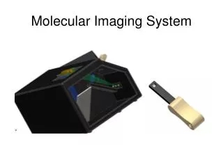

Molecular Imaging System
E N D
Presentation Transcript
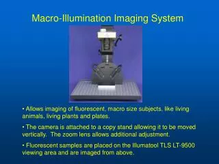
General Layout 600 – 900 nm light is used to illuminate fluorescent substances injected into the subject on the image plane.
Optigrid Functionality A portion of the OptiGrid at 200x y • Motors provide 120o phase shifts and focus • Grid moves on the x-axis 11.11 um to complete a phase change • Improves Clarity • Grid moves 2 mm on the z-axis to focus image • 30 line pair/mm on a 10x10 mm square (line pair approx .001 in wide) • Lines spatially modulate the illumination z x
Image Sharpening Provided by Qioptiq
3D Rendering Provided by Qioptiq
Subsystem Breakdown Imaging Unit Medical Testing Mechanical Software Optical Lens Mount Grid Mount Motor Control Image Capture Phantom Hand Image Rendering Lens System
Lens Selection Parameters • Focal Length • Magnification • Box Length = 250 mm • Box Height = 200 mm. • Lens size
Computar C mount • Alternative Lenses: • Telecentric Lens • Zoom Lens • Simple Lens
OSLO Simulation Simulation by Jeff
90 degrees Rear Projection 45 degrees
440mm Platen to Lens 3mm Lens to Grid
By Edmund NT56-35 by Edmund
250mm 250mm
Software System Capabilities • Consist of three separate programs • Kodak Molecular Imaging Software • Optigrid Motor Control GUI • MATLAB • Scripts will be developed as necessary for calibration and depth resolved discrimination • Optigrid Calibration • A system specific calibration process will be developed • Process will rely on manual coordination of three separate programs • Implement necessary algorithms to achieve depth discrimination, as described by Tromberg et al. in US Publication 2006/0184043
Objective: To mathematically investigate and invoke the necessary algorithms to achieve the depth discrimination, as described by Tromberg et al. in US Publication 2006/0184043 Bevilacqua, F., Cuccia, D., Durkin, A., Tromberg, B., “Quantitative analysis and imaging of subsurface heterogeneities using spatially structured illumination,” 2002.
Phantom Design • Objective: To develop a stable, long-term phantom whose optical properties can be predetermined and reproduced, at selected wavelengths, of human biological tissues of the hand, over a spectral range between 400 and 1,000 nm. • Purpose: • Simulate light distributions with a geometry of physical tissue. • Initially testing system designs • Calibration of optical devices. • Create reference measurements of reflective fluorescence. • How: • Understand of the key physical and biochemical characteristics of tissue that influence its interaction with light. • Match the absorption coefficient μa, scattering coefficient μs, and the anisotropic coefficient g(λ)
Design of Phantom • 3 components: • Casting material • Polyurethane • Absorbing material • phthalocyanin dye (Epolight 6084) • Scattering agent • Aluminum Oxide (alumina; Al2O3)
Total attenuation coefficient, as a function of concentration, of 0.954-mm-diameter micro-spheres.