Heart sound
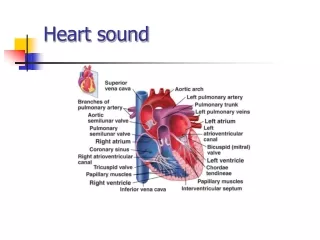
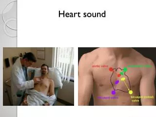
Heart sound. Area Of Auscultation. Area Of Auscultation. Pulmonary valve second intercostal space, left upper sternal border Aortic valve second intercostal space ,right upper sternal border Mitral valve fifth intercostal space , left midclavicular line

Heart sound
E N D
Presentation Transcript
Area Of Auscultation • Pulmonary valve second intercostal space, left upper sternal border • Aortic valve second intercostal space ,right upper sternal border • Mitral valve fifth intercostal space , left midclavicular line • Tricuspid valve fourth intercostal space, lower left sternal border
What we hear ? • We have all heard the heart make the usual sounds. LUB----------DUB • Lub is the first sound or S1 • Dub is the second heart sound or S2
Normal heart souands Normal
First heart sound S1 The “lub” in the lub – dub. • This sound is primarily because of the closing of the bicuspid and tricuspid valves. • Anatomically they are located between the atria and the ventricles • They close because the ventricles contract • The Pulmonic and Aortic valves are opening and blood is being forced into the arteries • Its maximum intensity is at the apex
S1 abnormalities • Loud S1 Mitral stenosis Tachycardia /hyperkinetic status • Soft S1 Mitral regurgitation Heart failure • Obesity • Shock • Pericardial effusion
Second heart sound S2 S2 is the “dub” in the lub- dub • The sounds are because of the closing of the Pulmonic and Aortic valves as the pressure from the arteries is greater then the pressure in the ventricles. • This is the end of systole
S2 components: • Has two components A2 and P2 • Inspiration decreases intrathoracic pressure, increases RV filling • RV is relatively weak, and an increase in filling results in slower emptying • Inspiration delays P2, causing audible splitting of S2 • P2 localized to pulmonary area while A2 audible all over the pericardium with max. intensity at aortic area
S2 abnormalities • Loud P2 Pulmonary hypertension • Soft P2 Pulmonary stenosis • Loud A2 Systemic hypertension • Soft A2 Aortic stenosis Aortic regurgitation
Split abnormalities • Wide splitting • Delay pulmonic closure: • RBBB • Pulmonary hypertension • Pulmonic stenosis • Early aortic closure: • MR • Fixed splitting Atrial septal defect • Reversed splitting • Lf bundle branch block • Sever aortic stenosis
Systole The time between the S1 and S2 sounds is: Lub------------Dub The ventricles contracting Blood flowing from the heart to the lungs and body Blood flowing across the Pulmonic and Aortic valves
Diastole The time between S2 and S1 is : Dub----------Lub The blood is flowing from the atria to the ventricles. The blood flowing across the bicuspid and tricuspid valves. The atrial contraction also occurs now
Third heart sound S3 • Is a low pitched early diastolic sound best heard with the bell at the apex. • also called ventricular gallop • Occure with rapid ventricular filling after the AV valves open. • It is best heard with the bell-side of the stethoscope at the apex of the heart • Causes • Normally in Children and during pregnancy • Pathological LVF MR
Fourth heart sound S4 • Low pitched sound occurs at late diastole due to atrial contraction if ventricles are non complaint. Just before S1 • Called a presystolic gallop or atrial gallop • It is always pathological • Causes: Hypertension Cardiomuopathy AS
Murmurs • These are abnormal sound and are longer duration s compared to heart sound produced due to the turbulence of blood flow through valves • Three Types: Systolic murmurs Diastolic murmurs Continuous machinary murmur