Download
1 / 55
550 likes | 908 Vues
Clinical Biochemistry Aspects of Cardiovascular Disease Dr Vivion Crowley MRCPath FRCPI Consultant Chemical Pathologist Biochemistry Department St James’s Hospital. Atherosclerosis is a major cause of morbidity and mortality. Clinically manifests as Coronary Heart Disease (CHD)

E N D
Clinical Biochemistry Aspects of Cardiovascular Disease Dr Vivion Crowley MRCPath FRCPI Consultant Chemical Pathologist Biochemistry Department St James’s Hospital
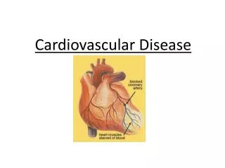
Atherosclerosis is a major cause of morbidity and mortality • Clinically manifests as • Coronary Heart Disease (CHD) • angina MI • Peripheral vascular disease (PVD) • Intermittent claudication limb amputation • Cerebrovascular disease • TIA Stroke
Atherosclerotic plaque is the key pathological lesion Underlying the morbidity and mortality associated with atherosclerosis
What are the risk factors for the development of atherosclerotic disease?
Other risk factors for atherosclerosis • Stress/Personality • Homocysteine • Lipoprotein (a) • Fibrinogen • Socioeconomic • Geographic • ? Depressive illness
How is‘obese’ defined? Body mass index (BMI)= weight/height2 (kg/m2) BMI 30 Health Hazard BMI 25 Healthy weight BMI 20 overweight Insufficient weight
25 USA Germany 20 UK 15 Netherlands 10 5 0 1980 1985 1990 1995 1998 Year Time trends in the prevalence of obesity (BMI > 30kg/m2) % WHO MONICA 1997 data , 1997
Central (Visceral) adiposity is associated with a greater risk of developing metabolic syndrome
Waist circumference is a clinically useful measure of central adiposity
Hypertension • Defined as BP ≥ 140/90 • Associated with stroke, CHD, Cardiac Failure, renal failure • Aetiology • Essential (primary HT) – polygenic disorder • Secondary HT (consider in younger hyepretensive) • Prevalence • - 33% White males • - 38% Black males
Secondary Hypertension • Renal disease • Renovascular disease (Renal artery stenosis) • Atheroma in older subjects • Fibromuscular dyspalsia in younger subjects • Coarctation of Aorta • Endocrine causes • Primary hyperaldosteronism (Conn’s syndrome) • Cushing’s Syndrome • Phaeochromocytoma • Renal tubular genetic defects • Liddle’s syndrome • Drugs • Streoids • OCP
Dyslipidaemia is a major risk factor for atherosclerosis • Dyslipidaemia refers to any perturbation in lipoprotein metabolism • -Hyperlipidaemia e.g. hypercholesterolaemia • Hypolipidaemia e.g. hypoalphalipoproteinaemia (low HDL) • The major lipoprotein particles • Very low density lipoprotein (VLDL) • VLDL remnant (IDL) • Low density lipoprotein (LDL) • High density lipoprotein (HDL)
What is the relationship of plasma lipids and CHD? • The plasma lipid profile consists of • Total Cholesterol (TC) • HDL Cholesterol (HDLC) • LDL Cholesterol (LDLC) • Triglycerides (TG) • TC:HDLC • Raised TC and LDLC levels are positively associated with CHD • HDLC levels are inversely associated with CHD • High level implies lower risk • Low level implies higher risk (M < 1.0mmol/L, F <1.3mmol/L) • Raised Triglyceride levels are independently associated with CHD
LDL cholesterol is calculated using the Friedewald formula Treatment targets for Plasma lipids TC <5.0mmol/L LDLC <3.0mmol/L (primary prevention) <2.5mmol/L (secondary prevention) HDL >1.0mmol/L in males >1.3mmol/L in females
Elevated Plasma Cholesterol levels are associated with increased CHD mortality
WHO Classification of Dyslipidaemia is now outdated • Adopted by WHO in 1970 • Based on laboratory parameters • Lipoprotein analysis • Lipoprotein electrophoresis • Serum/plasma appearance
Most practical classification takes account of aetiology and Plasma Lipid pattern Primary (Inherited) Secondary (Acquired)
Secondary Dyslipidaemias have multiple causes • Diabetes mellitus • Obesity • Alochol abuse • Hypothyroidism* • Nephrotic syndrome* • Chronic Renal failure* • Cholestasis* • PCOS • Drugs • Retinoic acid • Diuretics • Steroids • OCP • HAART • Cyclosporin * Predominant Hypercholesterolamia LFTs, U/E, TFTs, BMI, WC, Glycaemic status, medications and dietary habits need to be adequately assessed in the context of dyslipidaemia
Primary Dyslipidaemia should be considered in specific circumstances • Abnormal lipid profile without obvious secondary cause • Premature CVD • Family hx of Premature CVD • Family hx of dyslipidaemia • Identification of primary dyslipidaemia may have implications • CHD risk • Clinical management • Family screening • Genetic counselling
Primary dyslipidaemias can be sub-classified • Predominantly elevated plasma cholesterol • Polygenic hypercholesterolaemia • Monogenic hypercholesterolaemias e.g. FH, FDB • Predominantly elevated plasma triglyceride • Lipoprotein lipase (LPL) deficiency • ApoC-II deficiency • Familial hypertriglyceridaemia • Mixed (Combined elevated plasma Cholesterol and Trigs) • Familial combined hyperlipidaemia (FCH) • Dybetalipoproteinamia (Type III HPLA) • Very rare dylipidaemias • Low LDL syndromes e.g. abeta-, hypobeta-lipoproteinaemia • Low HDL syndromes • ApoA-I mutations • Tangier disease • LCAT deficiency • Miscellaneous – Lp(a), Hyperalphalipoproteinamia
Monogenic Hypercholesterolaemias • All known defective genes causing monogenic hyeprcholesteroloaemia • are involved in the receptor mediated uptake of LDL by LDL Receptor (LDLR)
Familial Hypercholesterolaemia (FH) is the most prevalent autosomal dominant inherited disorder Caused by mutation in the LDLR (Goldstein and Brown) High genetic heterogeneity (implications for genetic screening of populations) > 700 mutations • Heterozygous 1 in 500 • Homozygous/Compound Het 1 in 1,000,000
Biochemical Characteristics of FH • Pathogenesis • Reduction in functioning LDLR decreases plasma LDL catabolism • Also some degree of LDL overproduction • - ? increased IDL conversion or direct liver LDL overproduction • Lipid profile • Increased plasma Total Cholesterol 8-14mmol/L • Increased plasma LDL Cholesterol 6-11mmol/L • Normal or decreased plasma HDL Cholesterol • Normal plasma triglycerides • Lp(a) – may also be increased ( ? Role in increased CHD risk)
Clinical Characteristics of FH • Tendon Xanthomata are a pathognomic feature of FH • -Usual sites are extensor tendons on hands, Achilles tendon, pretibial tuberosity • Present in 70% Heterozgotes by 4th decade of life • Present in Homozygotes by age 5 years • Homozygotes also have cutaneous planar xanthomas • e.g. inter-digital spaces, buttocks, knees, hands
FH is associated with markedly increased risk of CHD and premature death • Heterozygotes • Mean age of onset of CHD is 43yrs (males) 53yrs (females) • Relative Risk (RR) was 8 pre-introduction of statins for Rx FH • RR in statin era is 3-4 • Homozygotes • Symptomatic CHD may be evident before age 10 years • Usually present by 20 yrs • Mean age of death from CHD is 26 yrs
Haemodynamically significant Aortic stenosis is a major cause of morbidity in Homozygous FH • Due to atheromatous involvement of the aortic root • Usually present by puberty • In Heterozygotes, aortic valve involvement is not characteristic
How do you diagnose FH? • Three established sets of diagnostic criteria • The Simon Broome FH Register • Dutch Lipid Clinic Network • US MEDPED Program
Differential diagnosis of FH • Polygenic Hypercholesetrolaemia • Familial Combined Hyperlipidaemia • Other monogenic Hypercholesterolaemia
Screening for FH • Universal Population screening – impractical, not cost effective • Screening within the clinical setting (Opportunistic screening) • Hyperchol, premature CHD, Fam Hx of CHD or dyslipidaemia • Cascade screening of FH relatives • Use diagnostic criteria (limited sensitivity) • Genetic approach -52-76% of patient who meet criteria are LDLR Mutation positive
Management of FH • Heterozygotes: • Effective lowering of LDL Chol can significantly reduce morbidity and mortality • Use of high dose Statins is the first line treatment • Statin may not adequately reduce LDL levels • Consider combination with Ezetimibe (18% further reduction), Resin or Fibrate • If lack of response consider LDL-apheresis + statin (rarely required now) • In females consider contraception if commencing statins or other lipid-lowering drugs • Regular non-invasive testing for silent ischemia (every 1-2 years depending on risk) • e.g. stress ECGs, thallium scans • Family screening is mandatory in FH
Dysbetalipoproteinaemia • Type III HPLA • Remnant particle disease • Pathophysiology: • Absence of ApoE R mediated removal of chylomicron and VLDL remnants • Mixed HPLA where plasma Cholesterol and Trigs are elevated to the similar levels • Mean untreated levels of P Chol and Trigs is 8-10mmol/L • Clinical features: Palmar xanthomatosis, tubero-euptive xanthomata • Associated with increased risk of premature CHD and PVD (approx 50%) • Excellent repsonse to Fibrates (± statin)
Genetics of Dysbetalipoproteinaemia There are several different genetically determined isoforms of ApoE • ApoE2/E2 is present in > 90% Type III HPLA • E2/E2 genotype frequency of 1 in 100 • However Type III HPLA prevalence is 1 in 5000-10000 • Further environmental “stresses” required to manifest this pheontype • e.g. T2DM, alcohol, hypothyroidism, obesity • Example of a gene-environment interaction • Other Mutations in ApoE e.g. R147W • -can cause an autosomal dominant form of Type III HPLA
CHD – clinical aspects • Spectrum of clinical presentation • Angina • Acute Coronary Syndrome (ACS) • Unstable angina MI • Symptoms of ACS • Severe crushing central chest pain • Dyspnoea • Cold sweat • Pallor • Nausea
Diagnosis of Acute Coronary Syndrome (ACS) • Clinical history • ECG • -STEMI or NSTEMI • -Q waves appear later • Clinical Biochemistry
“Older” Cardiac Biomarkers for Diagnosis of MI • Creatine Kinase (CK) • muscle enzyme • Nonspecific in that it may originate from skeletal or cardiac muscle • start to increase at 3-8h • Peak level 18-24h • Returns to normal 3-4 days • Aspartate transaminase (AST) • Found in Liver and muscle (an dother tissues) • Nonsepcific • Incraese 6-10h • Paek level 24h • Return to normal 3-5 days • Lactate dehydrogenase (LDH) • Nonspecific (LDH 1 isoform is more cardiospecific) • Peak at 72hrs • Return to normal 8-14 days
New Cardiac Biomarkers for ACS Diagnosis • CK-MB • Myocardium has higher concentration of CK-MB, more specific for heart • In ACS similar kinetics to total CK • CK-MB >6%of total CK indicates myocradial origin (Fractionated) • CK-MB mass >5 • Troponins • Regulatory complex in muscle consisting of 3 protein T, C, I • Increases in Troponin T or I are very specific for cardiac muscle damage • In ACS increase at 3-6 hr • Peak 18-24 hr • Can remain elevated for 7-10 days • A Troponin T or I taken at 12 hrs post onset of chest pain is very sensitive