Download
1 / 17
610 likes | 1.51k Vues
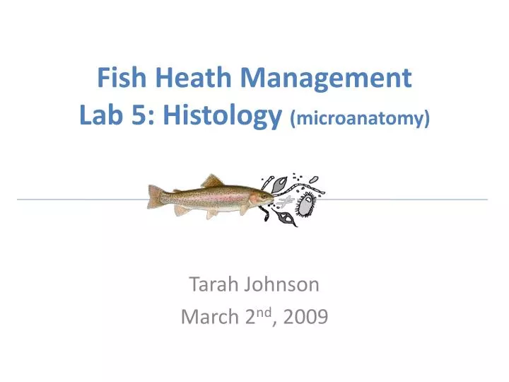
Fish Heath Management Lab 5: Histology (microanatomy). Tarah Johnson March 2 nd , 2009. Objectives. Be able to microscopically identify cells and structures in the liver, kidney, and gills

E N D
Fish Heath Management Lab 5: Histology (microanatomy) Tarah Johnson March 2nd, 2009
Objectives • Be able to microscopically identify cells and structures in the liver, kidney, and gills • Have the opportunity to microscopically examine histological sections of other fish organs (eyes, head, muscle, etc.)
Liver • Hepatocytes • Hepatic Sinusoid • Central Vein and or Hepatic Portal Vein • Bile Duct • Arteriole • Glycogen Vacuole
Central Vein and Hepatic Sinusoids Hepatic sinusoids Erythrocytes visible within the central vein 400x
400x 400x A Bile Duct Arteriole A. Glycogen Vacuole Erythrocytes visible within arteriole
Head Kidney • Interrenal gland • Chromaffin cell • Postcardinal vein • Hematopoietic tissue
400x A B C • Interrenal Gland • Chromaffin Cell • Postcardinal vein
40x A B A. Postcardinal vein B. Hematopoietic tissue
Trunk Kidney • Nephron (may not observe a complete nephron): Renal corpuscle, proximal tube, distal tubule, and collecting duct • Renal Corpuscle:Glomerulus, Bowman’s capsule, basement membrane of glomerulus • Melanocytes • Collecting duct and/or Mesonephric duct • Connective tissue
400x • Renal Corpuscle • Proximal Tubule • Distal Tubule C B A
400x • Glomerulus • Basement membrane of glomerulus • Bowman’s capsule C B A
400x C A B • Mesonephric Duct • Melanocytes • Connective tissue
Gills • Filament • Lamella • Goblet (or mucous) cell • Chloride cell • Pillar (or pilaster) cell • Gill filament cartilage
Lamella B C • Filament cartilage • Pillar cell • Chloride cell A 400x Gill filaments and lamellae 40x
400x Mucous or Goblet Cells
Laboratory Steps • Use proper microscope handling procedures (distributed in Lab 2) • Examine histology slides of liver, kidney, and gills • Examine additional histology slides of other organs
Be Sure to… • Clean oil immersion lenses • Clean slides and return to proper case (the same one you took it from) • Turn off microscopes and leave on low power objective when done