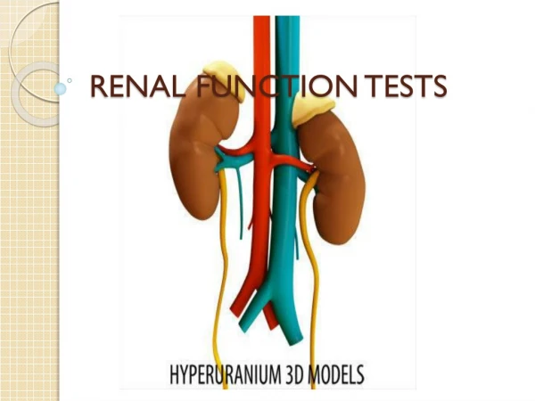

RENAL FUNCTION TEST
E N D
Presentation Transcript
RENAL FUNCTION TEST PRESENTED BY H GROUP Mohini, monika R, Ankita, Kinjal, Kinal, monika
INTRODUCTION: • Kidneys are the organ that filter waste products from the blood. • The kidneys serve three essential function: • They function as filter, removing metabolic product and toxins from the blood and excreting them through the urine. • They regulate the body’s fluid status, electrolyte balance and acid-base balance. • The kidney produce or activate hormones that are involved erythrogenesis, Ca²˖ metabolism and the regulation of blood pressure and blood flow.
Renal function may be assessed by measuring blood urea and serum creatinine. Renal function decreases with age , which must be taken into account when interpreting test values. • These tests primarly evaluate glomerular function by assessing the glomerular filtration • In many renal diseases, urea and creatinine accumulate in the blood because they are not excreted properly • These tests also aid in determining drug dosage for drugs excreted through the kidneys
What is Renal Function Test? • Renal function tests are use to detect the presence of renal diseases and assess their progress.
TESTS INVOLVED IN RFT: • Urea • Ammonia • Para Thyroid Hormone • Calcium • Uric acid • Potassium • Creatinine clearance • Glomerular filtration rate
CREATININE: • In the muscles creatine is converted to creatine phosphate which becomes the source of a high energy phosphate bond for the immediate reformation of ATP. • Creatinine is the byproduct of muscle energy metabolism and is produce at a constant rate according to the muscle mass of the individual. It is the substance that is easily excreted by the kidney. • By this method we can also estimate serum creatinine and urine creatinine.
Jaffe method Principle: In this colorimetric method creatinine reacts with picrate ion formed in alkaline medium to develop a red-orange colour. The colour produced from the sample is then compared in a colorimeter at wavelength of 520nm with that produced by known amount of creatinine under the same condition. • Creainine + picric acid →creatininepicrate (orange)
This divides into two types • Manual method and • Automated kinetic method The automated kinetic assay of creatinine is a method with the Jafee reaction but by using special type of spectrometer or autoanalyser.
Method: • To get clear supernatant, take 1ml of distilled water, serum, sodium tungstat and sulfuric acid. • Mix it well and centrifuge it for 5 min. • Label three tubes as blank, standard and test respectively.
Mix it well and incubate at room temperature for 10 min. • Take the OD against the blank at 520 nm. Calculation formula: Creatinine(mg/dL)= abs. of test/ abs. of std.*std. conc.
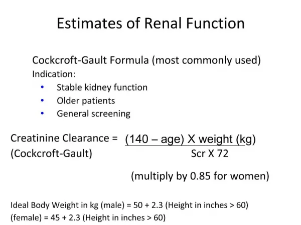
Creatinine Clearance • A measure of the amount of creatinine eliminated from the blood by the kidney. • The value is given in unit of millions per minute, representing the volume of blood cleared by the kidney per minute. • Calculation: C cr=U x V/P Where, U = Urine creatinine concentration in mg/dl, P = Serumecreatinine in mg/dl and V = Volume of urine in ml/mt.
Referance Range • Male: 0.7-1.3 mg/dL • Female: 0.4-1.1 mg/dL
Interpretation High creatinine level causes: • Acute and Chronic kidney disease • Ureter obstruction • Dehydration • Glomerulonephritis
PTH: PARATHYROID HORMONE PTH is a hormone secreted by the parathyroid gland There are four parathyroid glands located behind the thyroid
Role of PTH • To regulate calcium levels (in blood) • The parathyroid glands major function – regulate the calcium level in the body within a very narrow range (8.5 – 10.2 mg/dl)so that the nervous and muscular system can function properly. • Activation of vitamin D is very essential for calcium absorption from GI tract. • Vitamin D has to be converted into 1,25-dihydroxycholeciferol in the liver and kidney in the presence of PTH. Vitamin D1,25- dihydroxycholeciferol • PTH is also increased the formation of 1,25- dihydroxycholeciferol from 25-hydroxycholecalciferol.
PRINCIPLE OF THE TEST The DRG Intact PTH Immunoassay is a two-site ELISA [Enzyme-Linked ImmunoSorbent Assay] for the measurement of the biologically intact 84 amino acid chain of PTH.
Where, • Reagent 1= biotinylated PTH antibody • Reagent 2= peroxidase labeled PTH ab • Reagent A= saline with surfactant • Stop solution = 1N sulfuric acid • Referance range := 10-65 pg/ml
URIC ACID Introduction Uric acid is a naturally occuring waste product resulting from the breakdown of purine,crystalline compound found in certain foods. Under normal condition, uric acid dissolves in the blood, passes through the kidney and is eliminated with the urine.
Sometime the body produces too much uric acid or doesn’t filter out enough of it and that time uric acid level increase in blood. Main two condition are observed, 1. Hyperuricemia 2. Hypouricemia
URIC ACID BLOOD TEST • Introduction A uric acid blood test also known as a serum uric acid measurment, determine how much uric acid is present in your blood. The test can help determine how well your body produce and removes uric acid.
Intended use This reagent is for in vitro determination of uric acid in serum/plasma. • Clinical significance Uric acid is a metabolism of purines, nucleic acids and nucleo-proteins. Consequently, abno- rmal levels may be indicative of a disorder in the metabolism or in some genetic diseases.
Causes of increase and decrease of uric acid level - High level of uric acidin your blood can also indicate of a variety of conditions, inluding: - diabetes - gout (acute arthritis) - chemotherapy & radiation - leukemia(bone marrow disorders) - hypoparathyroidism - kidney disorders(stones) - multiple myeloma - metastasized cancer
- low levels of uric acid in the blood may inluding: - wilson’s disease - fanconi syndrome(cystinosis) - alcoholism - liver or kidney disease - a diet low in purines
Methodology 1. chemical method phototungstic acid method 2. enzymatic method The reagent is based on Trinder’s reaction, enzymatic and colorimetric method.
Raference range serum/plasma for women – 2.5-6.8mg/dl for men – 3.6- 7.7mg/dl
Limitations Limitations of uric acid testing are as follows: - Methodological interference and in cases of vitamin C, levodopa, and alphamethyldopa. - Early purine rich diet - several exercise increases uric acid - Rapid degradation of uric acid,which occurs at room temp.in the plasma of patients with tumor lysis syndrome.
UREA: • Urea is the chief nitrogenous waste of body. • Urea is the end product of protein metabolism. • After filtered by glomeruli. It is partilly reabsorbed by the renal tubules. Methodology: • Kinetic method • Enzymatic method
Principle: • Urea in the sample is hydrolizedenzymatically into ammonia(NH4+) and carbon dioxide (co2). • Ammonia ions formed reacts with α-ketoglutarate in a reaction catalysed by glutmatedehydrogenase(GLDH) with simultaneous oxidation of NADH to NAD+. Urease • Urea + 2H₂O 2NH₄⁺+ CO₃²⁻ GLDH • NH₄ ⁺ +2-Oxoglitarate +NADH L-glutamate +NAD ⁺+H₂O
The decrease in concentration of NADH, is proportional to urea concentration in the sample. ASSAY PROCEDURE: Mix well and aspirate standard followed by samples.
Calculation: Urea (mg/dl)=(∆ Abs of test/∆Abs of std)*conc. Of standard(mg/dl) REFERENCE VALUE: serum/plasma: 13-45mg/dl
Urea clearance test: • Urea clearance test is less than the GFR and it is influnced by the protein content of the diet. • Approximately 40% of filtered urea is normaly reabsorbed by tibules • The sensitivity of urea clearance is much less than the creatinine clearance because plasma concentration of urea is affected by number of factors. • Like, Dietary protien fluid intake infaction surgery, etc. • Nornaml value of urea clearance: 75% ml/min. • Urea clearance is defined as the volume(ml) of plasma that would be completely cleared of urea per minue.
It is calculated by the formula: Cm= U*V/P Cm= Maximum Urea clearance. U = Urea concentration in urine (mg/dl). V = Urine excreted per minute in ml. P = Urea concentration in plasma. • If the output of urine is more than 2ml per minute. • This is referred to as maximum urea clearance.
Standard Urea Clerance: • the urea clearance drastically changes when the volume of urine is less than 2ml/min. • This is known as standard urea clearance(C) and the and the normal value is around 54ml/min. Diagnostic importance: • A Urea clearance value below 75% of the normal is serious. Since it is an indicator of renal damage. • Blood urea level is found to increas only when the clearance falls below 50% normal. • Normal level of blood urea:20-40 mg/dl.
Causes for increasedblood urea: Pre-renal condition: -Dehydration: Severe vomiting, intestinal obstruction, diarrhea, diabetic coma, severe burns, fever and severe infections. Renal diseases: • Acute glomerulonephritis • Nephrosis • Malignant hypertension • Chronic pyelonephritis
Decreased blood urea: • Urea concentration in serum may be low in late pregnancy, in starvation, in diet grossly deficient in protein and in hepatic failure. • Azotemia:= • Increase in the blood level of NPN(creatinine,urea, uric acid) is referred to as azotemia and is the hallmark of kidney failure.
Calcium: • Calcium plays an important role in: • nerve impulse transmission, • muscle contraction, • pancreatic insulin release, • as a core factor for some enzyme reactions and blood coagulation, • and most important bone and tooth structural integrity. Normal total calcium values 8.8‐10.3 mg/dl.
Hypocalcemia usually implies a deficiency in either the production or response to parathyroid hormone(PTH) or vitamin D • Hypercalcemiais an increased calcium concentration and it is usually associated with malignancy or metastatic diseases.
Reference ranges in males • Younger than 12 months : not established • Age 1-14 yr : 9.6-10.6 mg/dl • Age 15-16 yr : 9.5-10.5 mg/dl • Age 17-18 yr : 9.5-10.4 mg/dl • Age 19-21 yr : 9.3-10.3 mg/dl • Age 22 yr and older : 8.9-10.1 mg/dl
OCPC Method • Principle: calcium in an alkaline medium combines with o – Cresolphthalein complexone to form a purple coloured complex. Intensity of the colour formed is directly proportional to the amount of calcium present in the sample. • Calcium + OCPC Purple coloured complex
Procedure • Wavelength/filter: 570nm(Hg 578 nm)/yellow • Temperature : R.T. • Light path : 1 cm • Pipette into clean dry test tubes labelled as blank(B),standard(S), and Test(T)
Addition sequence B S T (ml) (ml) (ml) Buffer Reagent(L1) ColourReagent(L2) Distilled water Calcium Standard (S) Sample • 0.5 0.5 0.5 • 0.5 0.5 0.5 • 0.02 - - • 0.02 - • - - 0.02 • Mix well and incubate at R.T.(25 ℃) for 5 min. Measure the absorbance of the standard(Abs.S), and test sample(Abs.T) against the blank,within 60 min.
Calculations • Calcium in mg/dl = Abs.T ______ X 10 Abs.S
Arsenazo iii method : • Principle : Calcium combines specifically with Arsenazo iii at a neutral pH to forma blue purple coloured complex. Intensity of the colour formed is directly proportional to the amount of calcium present in the sample. • Calcium + Arsenazo iii Blue purple coloured complex