Chap 12 Cell Cycle
Chap 12 Cell Cycle. 100 µm. 200 µm. 20 µm. (a) Reproduction. An amoeba, a single-celled eukaryote, is dividing into two cells. Each new cell will be an individual organism (LM).

Chap 12 Cell Cycle
E N D
Presentation Transcript
100 µm 200 µm 20 µm (a) Reproduction. An amoeba, a single-celled eukaryote, is dividing into two cells. Each new cell will be an individual organism (LM). (b) Growth and development. This micrograph shows a sand dollar embryo shortly after the fertilized egg divided, forming two cells (LM). (c) Tissue renewal. These dividing bone marrow cells (arrow) will give rise to new blood cells (LM). Figure 12.2 The functions of cell division
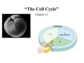
0.5 µm A eukaryotic cell has multiplechromosomes, one of which is represented here. Before duplication, each chromosomehas a single DNA molecule. Chromosomeduplication(including DNA synthesis) Once duplicated, a chromosomeconsists of two sister chromatidsconnected at the centromere. Eachchromatid contains a copy of the DNA molecule. Centromere Sisterchromatids Separation of sister chromatids Mechanical processes separate the sister chromatids into two chromosomes and distribute them to two daughter cells. Centrometers Sister chromatids Figure 12.4 Chromosome duplication and distribution during cell division
G1 M phase S G2
If a cell stops in the cell cycle it is in the G0 stage. Many mammal cells, such as this neuron or muscle cells, remain permanently or semipermanently in G0.
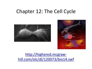
Mitosis and Cytokinesis Animation • http://www.sumanasinc.com/webcontent/animations/content/mitosis.html • http://highered.mcgraw-hill.com/olc/dl/120073/bio14.swf
PROMETAPHASE G2 OF INTERPHASE PROPHASE Centrosomes(with centriole pairs) Aster Fragmentsof nuclearenvelope Early mitoticspindle Kinetochore Chromatin(duplicated) Centromere Nonkinetochoremicrotubules Kinetochore microtubule Chromosome, consistingof two sister chromatids Nuclearenvelope Plasmamembrane Nucleolus Figure 12.6 Exploring The Mitotic Division of an Animal Cell
Prometaphase • The nuclear envelope fragments. • The microtubules of the spindle can now invade the nuclear area and interact with the chromosomes, which have become even more condensed. • Microtubules extend from each centrosome toward the middle of the cell. • Each of the two chromatids of a chromosome now has a kinetochore, a specialized protein structure located at the centromere. • Some of the microtubules attach to the kinetochores, becoming “kinetochore microtubules.” These kinetochore microtubules jerk the chromosomes back and forth. • Nonkinetochore (polar) microtubules interact with those from the opposite pole of the spindle. • G2 of Interphase • A nuclear envelope bounds the nucleus. • The nucleus contains one or more nucleoli (singular, nucleolus). • Two centrosomes have formed by replication of a single centrosome. • In animal cells, each centrosome features two centrioles. • Chromosomes, duplicated during S phase, cannot be seen individually because they have not yet condensed. • The light micrographs show dividing lung cells from a newt, which has 22 chromosomes in its somatic cells (chromosomes appear blue, microtubules green, intermediate filaments red). For simplicity, the drawings show only four chromosomes. • Prophase • The chromatin fibers become more tightly coiled, condensing into discrete chromosomes observable with a light microscope. • The nucleoli disappear. • Each duplicated chromosome appears as two identical sister chromatids joined together. • The mitotic spindle begins to form. It is composed of the centrosomes and the microtubules that extend from them. The radial arrays of shorter microtubules that extend from the centrosomes are called asters (“stars”). • The centrosomes move away from each other, apparently propelled by the lengthening microtubules between them.
METAPHASE ANAPHASE TELOPHASE AND CYTOKINESIS Metaphaseplate Cleavagefurrow Nucleolusforming Nuclear envelopeforming Daughter chromosomes Centrosome at one spindle pole Spindle
Anaphase • Anaphase is the shortest stage of mitosis, lasting only a few minutes. • Anaphase begins when the two sister chromatids of each pair suddenly part. Each chromatid thus becomes a full- fledged chromosome. • The two liberated chromosomes begin moving toward opposite ends of the cell, as their kinetochore microtubules shorten. Because these microtubules are attached at the centromere region, the chromosomes move centromere first (at about 1 µm/min). • The cell elongates as the nonkinetochore microtubules lengthen. • By the end of anaphase, the two ends of the cell have equivalent—and complete—collections of chromosomes. • Metaphase • Metaphase is the longest stage of mitosis, lasting about 20 minutes. • The centrosomes are now at opposite ends of the cell. • The chromosomes convene on the metaphase plate, an imaginary plane that is equidistant between the spindle’s two poles. The chromosomes’ centromeres lie on the metaphase plate. • For each chromosome, the kinetochores of the sister chromatids are attached to kinetochore microtubules coming from opposite poles. • The entire apparatus of microtubules is called the spindle because of its shape. • Telophase • Two daughter nuclei begin to form in the cell. • Nuclear envelopes arise from the fragments of the parent cell’s nuclear envelope and other portions of the endomembrane system. • The chromosomes become less condensed. • Mitosis, the division of one nucleus into two genetically identical nuclei, is now complete. • Cytokinesis • The division of the cytoplasm is usually well underway by late telophase, so the two daughter cells appear shortly after the end of mitosis. • In animal cells, cytokinesis involves the formation of a cleavage furrow, which • pinches the cell in two.
Aster Centrosome MetaphasePlate Sisterchromatids Kinetochores Overlappingnonkinetochoremicrotubules Kinetochores microtubules 0.5 µm Microtubules Chromosomes Centrosome 1 µm Figure 12.7 The mitotic spindle at metaphase
Pearson Mitosis Lab Print out answers to Lab Quiz 1 tonight
Onion Mitosis Click Here for Onion Mitosis Site
Prophase Interphase
Metaphase Telophase
Metaphase Anaphase
Prophase Metaphase Telophase Anaphase
Something from the active Mitotic cell triggers the G1 cell to undergo mitosis too
MPF is only active when there is a high cyclin concentration Animation MPF will phosphorylate many enzymes - which will move the cell past the G2 checkpoint into M phase - it will also make a proteolytic enzyme that destroys cyclin and thus turn off MPF Cdk and Cyclin combine to form MPF - which is an active from of Cdk
Regulation of Cell Cycle • Internal regulation- ex: APC anaphase promoting complex is inactive until all kinetocore are attached • External Regulation- when cells are not touching then PDGF is released to promote cell division
3 2 1 EXPERIMENT Scalpels A sample of connective tissue was cut up into small pieces. Petriplate Enzymes were used to digest the extracellular matrix,resulting in a suspension of free fibroblast cells. Cells were transferred to sterile culture vessels containing a basic growth medium consisting of glucose, amino acids, salts, and antibiotics (as a precaution against bacterial growth). PDGF was added to half the vessels. The culture vessels were incubated at 37°C. Without PDGF With PDGF Figure 12.17 Inquiry Does platelet-derived growth factor (PDGF) stimulate the division of human fibroblast cells in culture?