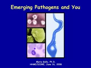
The Role of Clinical Microbiologists in Combatting Emerging Pathogens
This article discusses the vital role of clinical microbiologists in diagnosing and managing infectious diseases. It highlights the educational journey of Dr. Peter H. Gilligan, detailing his extensive experience in microbiology and public health. The piece emphasizes the contribution of microbiologists to public health, outbreak investigations, and the challenges they face, including governmental oversight and financial pressures. The text also outlines how to pursue a career in clinical microbiology, including educational requirements and career prospects in various industries.


The Role of Clinical Microbiologists in Combatting Emerging Pathogens
E N D
Presentation Transcript
Emerging pathogens 2007 • Peter H. Gilligan PhD • Clinical Microbiology-Immunology Labs • UNC Hospitals
How I became a clinical microbiologist • Obtained doctoral degree in microbiology at the University of Kansas • Did post-doctoral training (2 years) in medical and public health microbiology at UNC Hospitals • Director of Microbiology Labs at St Christopher’s Hospital for Children (Philadelphia) for 4 years • Past 20+ years, Associate Director then Director of the Clinical Microbiology-Immunology Labs at UNC Hospitals • Have served on medical school admission committee for approximately 15 years and the MD/PhD advisory (admissions) committee for the past 10 years
What do clinical microbiologists do? • We serve: • our patients • our health care-providing colleagues, physicians, nurses, physician assistants, pharmacy colleagues • hospital administrators • We make money for the institution • general public by insuring the public health • Involved in studying outbreaks of several emerging infectious diseases
How do we serve? • central role in the diagnosis and management of infectious diseases • central role in infection control and antimicrobial use • recognize emerging disease threats and outbreaks including bioterrorism events • we educate & train health care providers • we create new knowledge (research) to deal with practical problems
Best things about my job • Direct impact on patient care and public health of the community • Intellectually challenging job requiring a broad fund of knowledge-need to know a little about a lot of things –I am never bored!!!!!!! • Work with highly motivated and intelligent individuals • Get to be at the cutting edge of infectious disease diagnosis
Worst things about my job • Incredible amounts of governmental oversight • Increasing emphasis on financial aspects of the job • Declining talent pool of technologists • Need to be responsible for an organization that run 24/7/365-we never close. Personally have worked through ice storms, blizzards, and hurricanes.
How you can become a clinical microbiologist • CLS programs available here, ECU, WCU, WSSU, Wake Forest, UNC-CH • Education is also available on line • 2 more years of school to get a BS in CLS • Take ASCP certification exam to become certified as a MT. • Starting salary is 38,000 and up • Career options are amazingly diverse; many former UNC students work in leadership positions in the pharmaceutical and biotech industries
HIV* Avian influenza SARS* Cryptosporidium* E. coli O157:H7* Nipah virus nv Creutzfeldt-Jakob disease Sin Nombre Virus West Nile Virus Clostridium difficle* Bacillus anthracis(BT agent) Cyclospora CA-MRSA* Rapidly growing mycobacterium* Rotavirus* BK virus* Chlamydia pneumoniae Pencillinum marneffei Legionella* MDR- TB and pneumococcus* Burkholderia cepacia complex* VRE/VRSA* Helicobacter pylori* Invasive Group A streptococcal disease* HHV-6* HPV* HCV* Emerging/Re-emerging Infectious Diseases in the past 25 years
How do new pathogens emerge • Organisms that jump species barriers • Changing ecosystems • Changes in food production techniques • Evolution of medical devices and care • Long term survival of immunosuppressed • Pathogens that are detected because of new technology • Misuse of micro-organisms • Biocrime/bioterrorism • Organism evolution as a result of human intervention • Antibiotic pressure
How do microbes change? • Bacteria, because they evolve very quickly, can readily adapt to hostile environments • Assume a generation time for a bacteria of 50 minutes • 30 generations/day; or 220,000 bacterial generations for each human generation (assume generation is 20 years) • Bacteria have a huge evolutionary advantage over humans
How emerging pathogens develop? • Mutation drives evolution • constantly occurring • usually silent or lethal • environmental pressure such as antibiotics may select “resistance” mutation • Key feature of success of antibiotic resistant strains is their genetic fitness I.e. their ability to compete in a complex microbial environment • Recognition that certain bacteria may be hypermutators because of mutation in DNA repair genes • These strains may not be as “fit” as wild-types but may predominant in certain chronic infections such as P. aeruginosa causing chronic pulmonary infections in CF patients
How do emerging pathogens develop? • Recombination • Resistance genes from antibiotic producing organisms • genetic exchange of resistant genes can occur among organisms which are genetically diverse • Think Cholera toxin genes to E. coli • transfer of resistance/virulence genes can be mediated by plasmids/phage/transposons/ integrons
Organisms that jump species barriers • HIV, SARS, Avian flu • HIV likely jumped from primates to humans • SARS from pigs(?) • Avian flu-hasn’t yet made the jump from birds to humans because human to human spread is rare, if it occurs at all. However mutation may result in that occurring. • Technology allows us to quickly develop diagnostics for new pathogens • Took years to develop HIV diagnostics • Took weeks to develop SARS diagnostics
Changing ecosystems • Lyme disease • A perfect storm • Farmland in New England returned to forest • Natural predators for deer were eliminated • Deer populations and the ticks they carried increased because of ecosystem changes • People built homes and spent increasing amounts of time in the woods • This resulted in increased exposure to deer ticks that carried Borrelia • Ticks were pencil point in size and often difficult to see
Changes in food production techniques • Increased use of factory farming • Feedlots bring together large numbers of animals who produce large amounts of waste • Waste can lead to run-off of EHEC that can contaminant adjacent fields as was seen in recent spinach outbreaks • Large meat packing operations can result in 50 ton lots of ground meat containing 100s of animals • Meat can be distributed throughout the US • Contaminated lots can then lead to large scale outbreaks
Changes in medical care • Immunosuppression either as a result of HIV or medically therapy (ex. transplants) results in emerging infections • Pneumocystis, MAC, toxoplasma and CMV in HIV patients • CMV, adenovirus and HHV-6 in transplant patients • The use of indwelling artificial materials such as catheters, shunts, artificial joints present new ecosystems and new organisms • Examples-coagulase negative staphylococci growing as a biofilm on artificial joints/catheters/shunts • Rapidly growing mycobacteria causing keratitis following LASIK surgery
Pathogens detected with new technology • Prime example is HCV • Viral genome elucidated using molecular cloning techniques • Broad range 16S RNA primers are used to detect non-cultivable bacteria • Next big thing- application of molecular tools to understand how mixed microbial populations cause disease • Likely diseases caused by mixed microbial populations are bacterial vaginosis, peridontal disease, inflammatory bowel disease, CF lung disease
How does bacterial resistance develop? • Bacterial resistance develops in response to antimicrobial pressure • It is estimated that 3 million lbs of antimicrobials are used each year in the US • Much of it is used in children to treat viral respiratory illness • Estimated that 3/4 of children in US younger than two receive antimicrobials • Children then may serve as a key role for the emergence of antimicrobial resistance • 10x that amount are used in animals • End result- tremendous selective pressure that results in the emergence of bacterial resistance
UNC-ED • 6% of wounds from ED in 1st quarter of 2005 grew MRSA • 45% of wounds from ED in 2nd quarter of 2005 grew MRSA • ? Due to proliferation of CA-MRSA? • GOAL • To characterize and determine the prevalence of CA-MRSA isolates at UNC hospitals
Molecular analysis: CA- vs. HA-MRSA Adapted from Weber, CID, 2005:41S
Virulence of CA-MRSA • Panton-Valentine leukocidin (PVL) • Hemolysin first reported in 1932 by Panton and Valentine • Located on mobile phage • 2 co-transcribed genes, lukS-PV and lukF-PV • The two subunits form a hexameric pore-forming cytolytic toxin with a high affinity for PMNs and macrophages • PVL producing strains associated with skin and soft tissue infections and necrotizing pneumonia • Rarely associated with osteomyelitis, septicemia, or endocarditis • Rare HA-MRSA strains with PVL have similar clinical syndrome • Usually only 2% of all S. aureus isolates produce PVL but found in the majority of epidemic CA-MRSA strains
SCCmec types (Staphylococcal chromosome cassette) 21-24 Adapted from Diederen and Kluytmans, JID, 2005
Susceptibility Patterns tmp-smz ery vanc gent pen clinda cefox ery vanc doxy doxy gent clinda cefox tmp-smz pen CA-MRSA HA-MRSA 93% are erythromycin resistant 16% clindamycin resistant
CA-MRSA Timeline Children without identifiable risk factors Prison and jail populations 2003 Late 1990s 1980 Mid 1990s 2000 Necrotizing pneumonia, United States and Europe Outbreak in Detroit 2/3 of patients were IVDU 1998 - Athletes/sports teams 1999 - Native Americans IVDU=intravenous drug users Naimi TS et al. JAMA. 2003;290:2976-2984. Zetola N et al. Lancet Infect Dis. 2005;5:275-286. Levine DP et al. Ann Intern Med. 1982;97:330-338. CDC. Morb Mortal Wkly Rep. 2003;52:793-795. Groom AV et al. JAMA. 2001;286:1201-1205. Herold BC et al. JAMA. 1998;279:593-598. CDC. Morb Mortal Wkly Rep. 2001;50:919-922. Gillet Y et al. Lancet. 2002;359:753-759. CDC. Morb Mortal Wkly Rep. 1999;48:707-710.
Clinical presentation CA-MRSA • CA-MRSA • SSTIs (abscesses, cellulitis, folliculitis, impetigo, furunculosis*) • Typically treated with excision and drainage; +/- oral antibiotics • Occasionally require IV antibiotics, hospitalization and surgical intervention • Necrotizing pneumonia especially in young people secondary to influenza was reported this flu season • Mortality was over 50%- median time to death 3.5 days • Median age was 17.5 years • 5 isolates from Louisiana were CA-MRSA genotype of the same PFGE type • Both levofloxacin and inducible clindamycin resistance seen in these isolates
Case 5 • The patient is a 16 yo who presents with shoulder and left chest wall pain • An MRI is ordered because of concerns about a abscess • The patient becomes hypotensive, SOB, is intubated and admitted to the MICU. • Prior to admission, he denied fever, chills, cough and night sweats • He lives on a farm in rural central NC with exposures to dogs cats and horses • In the past year a horse had been put done due to “strangles.” Strangles is a respiratory infection caused by Streptococcus equi
Case 5 • No contributory travel or sexual history. Does not use drugs or alcohol • Two months previously he had a right-sided preauricular abscess incised and drained • Treated with Augmentin and infection resolved • On PE, afebrile, pulse was 103 bpm, RR 30 and BP 99/62 • Skin examination was significant for a small violaceous lesion at the site of the prior abscess • Had several pustules on his leg and a hyperpigmented macules on his left great toe • LDH was highly elevated, he was anemic and had a sed rate of 60
Study Design I. Outpatient wound cultures (SSTIs) with MRSA (6/05 to 3/06), n=233 • Nosocomial MRSA isolates (blood) (6/05 to 4/06), n=76 • Wound cultures with MRSA regardless of location (6/06-7/06), n=100 • Respiratory cultures with MRSA from Cystic Fibrosis (CF) patients • (10/05 to 4/07), n=339 V. Child care centers VI. All isolates recovered at Lilongwe Medical Center (6-06-2-07) n>100 • Definition of CA-MRSA Panton-Valentine leukocidin positive SCCmec type IV
IV II 500 I. PVL and SCCmec Characterizationof outpatient wound isolates SCCmec typing** 72% 0.5% 0.5% 11% 9% 6% n=233 (n= 42) (n= 191) ** Oliveira and Lencastre (2002) Antimicrob Agents Chemother46, 2155-61.
Study Design I. Outpatient wound cultures (SSTIs) with MRSA (6/05 to 3/06), n=233 • Nosocomial MRSA isolates (blood) (6/05 to 4/06), n=76 • Wound cultures with MRSA regardless of location (6/06-7/06), n=100 • Respiratory cultures with MRSA from Cystic Fibrosis (CF) patients • (10/05 to 4/07), n=339 V. Child care centers VI. All isolates recovered at Lilongwe Medical center (in June 07) • Definition of CA-MRSA Panton-Valentine leukocidin positive SCCmec type IV
SCCmec typing IV II 500 II. PVL and SCCmec Characterizationnosocomial blood isolates # of isolates 55% 21% 15% 9% n=76 (n= 16) (n= 60)
Clinical Characterization CA HA I CA Molecular Characterization 1 14 1 16 HA 0 41 1 42 I 0 1 17 18 2 72 2 II. Clinical and Molecular Analysisnosocomial blood isolates N=76
Study Design I. Outpatient wound cultures (SSTIs) with MRSA (6/05 to 3/06), n=233 • Nosocomial MRSA isolates (blood) (6/05 to 4/06), n=76 • Wound cultures with MRSA regardless of location (6/06-7/06), n=100 • Respiratory cultures with MRSA from Cystic Fibrosis (CF) patients • (10/05 to 4/07), n=339 V. Child care centers VI. All isolates recovered at Lilongwe Medical center (in June 07) • Definition of CA-MRSA Panton-Valentine leukocidin positive SCCmec type IV
IV II 500 III. PVL and SCCmec Characterizationof 2006 wound isolates SCCmec typing 72% # of isolates 9% 10% 8% n=100 (n= 81) (n= 19)
Thanks to: • Melissa Miller • Jennifer Goodrich • Joel Wedd • Mwai Makoka and the UNC project Lilongwe • Tameaka Sutton-Shields • Kyle Rodino • All the CMIL technologists who identify, save and freeze isolates so we can do this research