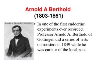
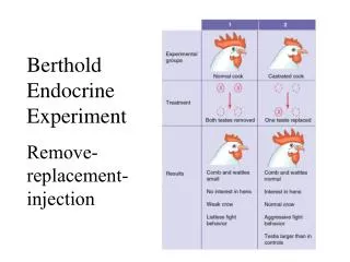
Berthold Endocrine Experiment Remove- replacement-injection
Berthold Endocrine Experiment Remove- replacement-injection. Objectives. 1. Chemical classes of hormones 2. Biosynthesis of a particular hormone 3.Transport of the hormones 4.Recognition & signaling of the hormone 5.Functions of the hormones . 6.Degradation of the hormone.

Berthold Endocrine Experiment Remove- replacement-injection
E N D
Presentation Transcript
Berthold Endocrine Experiment Remove- replacement-injection
Objectives 1. Chemical classes of hormones 2. Biosynthesis of a particular hormone 3.Transport of the hormones 4.Recognition & signaling of the hormone 5.Functions of the hormones. 6.Degradation of the hormone.
Endocrine Methods • Remove-replacement-injection • Purification and cloning • Synthesis and production of hormone • Test biological activity with pure hormones • Development of antibodies • Localization by immunocytochemistry • Establish assays (RIA) • Microarray,deep sequencing, proteomics • Knock-out/Knock down/mutants
Hormone Types/Functions Three structural divisions: 1) Amines--H2O sol. (small--AA) catecholamines and thyroid hormones 2) Steroids--lipid sol. cyclic hydrocarbon derivatives from cholesterol 3) Peptide/protein -- H2O sol. largest, complex
Amines • Hormones derived from tyrosine and tryptophan. • Include hormones secreted by adrenal medulla, thyroid, and pineal glands.
Thyroid Hormones • Tyrosine derivatives bound together. • Contain 4 iodine atoms (T4). • Contain 3 iodine atoms (T3). • Small, non-polar molecules. • Soluble in plasma membranes.
Steroids • Lipids derived from cholesterol. • Are lipophilic hormones. • Testosterone • Estradiol • Cortisol • Progesterone
Peptides/Proteins • Chains of amino acids (< 100 amino acids in length). • ADH • Insulin • Long polypeptides (>100) bound to one or more carbohydrate groups. • FSH • LH
Biosynthesis of Peptides and Protein Hormones DNA (The gene) RNA (Primary transcript) RNA processing mRNA translation Pre-(Pro)-hormone Proteolysis via signal peptide cleavage Pro-hormone Proteolysis via second modification Glycosylation phosphorylation Hormone
Proopiomelanocortin (POMC) gene 5’ 3’ 5’ 3’ mRNA 1 2 3 4 5 6 7 C N Signal peptide Products in corticotrophic cell of the anterior pituitary 4 6 7 ACTH b-lipotropin Products in the intermdeiary gland 6 8 a-MSH CLIP g-lipotropin g-MSH b-endorphin b-MSH MET-enkephalin
Classification of chemical communication systems 1) Autocrine--secretion that affects the same cell which the secretion originated Ex: Adrenergic nerve endings 2) Paracrine--secretion that affects neighboring cells Ex: Inflammatory response 3) Endocrine--a secretion of a chemical substance that is released into the blood and affects a distant target
4) Exocrine--secretion of a substance that is released onto surface of animal--including internal structures
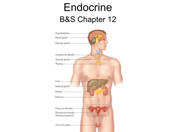
Exocrine Glands/tissues: --possess ducts --salivary glands, intestinal epithelium, secretory cells in stomach, and secretory cells of the liver and pancreas Endocrine Glands/tissues: --lack a definite duct --Adrenal gland, GI tract, heart, kidney, ovary, pancreas, thyroid, pituitary, placenta, testes, and thymus
Hypothalamus Bone Hypothalamus Optic chiasm Connecting stalk (infundibulum) Anterior lobe of pituitary (b) Posterior lobe of pituitary Posterior pituitary Anterior pituitary (a) Fig. 7-8, p.265
Posterior Pituitary • Also called the neurohypophysis. • Formed by down growth of the brain during fetal development. • Is in contact with the infundibulum. • Nerve fibers extend through the infundibulum.
Anterior Pituitary • Master gland (also called adenohypophysis). • Derived from a pouch of epithelial tissue that migrates upward from the mouth. • Consists of 2 parts: • Pars distalis: anterior pituitary. • Pars tuberalis: thin extension in contact with the infundibulum.
Hypothalamic Control of Posterior Pituitary • Hypothalamus produces: • ADH: supraoptic nuclei. • Oxytocin: paraventricular nuclei. • Hormones transported along the hypothalamo-hypophyseal tract. • Stored in posterior pituitary. • Release controlled by neuroendocrine reflexes.
Posterior Pituitary (neurohypophysis) --releases neurohormones 1) antidiuretic hormone (vasopressin) 2) oxcytocin
Hypothalamic Control of the Anterior Pituitary • Hormonal control rather than neural. • Hypothalamus synthesizes releasing hormones and inhibiting hormones. • Hormones are transported to axon endings of median eminence. • Delivers blood and hormones to anterior pituitary via portal system.
Hypothalamic Control of the Anterior Pituitary • Hormones secreted into the hypothalamo-hypophyseal portal system regulate the secretions of the anterior pituitary.
Feedback Control of the Anterior Pituitary • Anterior pituitary and hypothalamic secretions are controlled by the target organs they regulate. • Negative feedback inhibition by target gland hormones.
Feedback Control of the Anterior Pituitary • Negative feedback at 2 levels: • The target gland hormone can act on the hypothalamus and inhibit releasing hormones. • The target gland hormone can act on the anterior pituitary and inhibit response to the releasing hormone.
Water vs. Lipid Water soluble Lipid soluble hydrophilic hydrophobic external (2nd mess.) internal external receptors cytoplasmic rec. short half-life long half-life intermediary resp. Long-term resp. protein activation gene activation
Mechanisms of Hormone Action • Hormones of same chemical class have similar mechanisms of action. • Location of cellular receptor proteins. • Target cell must have specific receptors for that hormone (specificity). • Hormones bind to receptors with high bond strength (affinity). • Low capacity of receptors (saturation).
Hormones That Bind to Nuclear Receptor Proteins • Lipophilic steroid and thyroid hormones bound to plasma carrier proteins. • Hormones dissociate from carrier proteins to pass through lipid component of the target cell membrane. • Receptors for the lipophilic hormones are known as nuclear hormone receptors.
Nuclear Hormone Receptors • Function within cell to activate genetic transcription. • mRNA directs synthesis of specific enzyme proteins that change metabolism. • Receptor must be activated by binding to hormone before binding to specific region of DNA called HRE (hormone responsive element). • Located adjacent to gene that will be transcribed.
Mechanisms of Steroid Hormone Action • Steroid receptors located in cytoplasm. • Bind to steroid hormone. • Translocates to nucleus. • DNA-binding domain binds to specific HRE of the DNA. • Dimerization occurs. • Stimulates transcription.
Mechanism of Thyroid Hormone Action • Receptor proteins located in nucleus. • T3 binds to ligand-binding domain. • DNA-binding domain can then bind to the half-site of the HRE. • Other half-site is vitamin A derivative 9-cis-retinoic acid. • Two partners can bind to the DNA to activate HRE.
Hormones That Use 2nd Messengers • Cannot pass through plasma membrane. • Catecholamines, polypeptides, and glycoproteins bind to receptor proteins on the target cell membrane. • Actions are mediated by 2nd messengers (signal-transduction mechanisms). • Extracellular hormones are transduced into intracellular second messengers.
Hormones That Use 2nd Messengers 2nd messenger systems: • Adenylate cyclase • Phospholipase C • Tyrosine kinase • NO • various kinases
Adenylate Cyclase-cAMP • Hormone binds to receptor protein. • Dissociation of a subunit of G-protein. • G-protein binds and activates adenylate cyclase. • ATP cAMP + PPi • cAMP attaches to inhibitory subunit of protein kinase.