Download
1 / 17
220 likes | 585 Vues
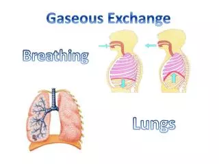
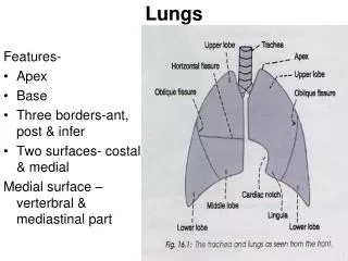
Lobes of the Lungs. Lungs are divided into _______ (in most species) Pattern varies with species Dogs, cats, cattle, pigs, goats, sheep → deeply fissured into lobes L: Cranial and Caudal R: Cranial, Middle, Caudal, Accessory Horses → least subdivided L: All one lobe

E N D
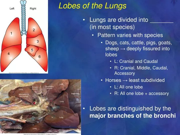
Lobes of the Lungs • Lungs are divided into _______ (in most species) • Pattern varies with species • Dogs, cats, cattle, pigs, goats, sheep → deeply fissured into lobes • L: Cranial and Caudal • R: Cranial, Middle, Caudal, Accessory • Horses → least subdivided • L: All one lobe • R: All one lobe + accessory • Lobes are distinguished by the major branches of the bronchi
Lungs continued • __________- small, well-defined area on medial side of the lung • Location of air, blood, lymph, and nerves entering and leaving the lung.
Pulmonary Circulation- THIS IS A REVIEW!!!! • ______________ blood enters the lungs from ______ ventricle of heart through the pulmonary ______. • Pulmonary artery splits into left and right pulmonary arteries that enter the two lungs • Pulmonary arterioles enter capillary networks around the alveoli • Oxygenated blood returns to the left atrium in the pulmonary veins.
Thoracic Cavity • Bound by __________ vertebrae dorsally, ______ & _____________ muscles laterally, the __________ ventrally, and the _____________caudally. • Mediastinum – area between lungs REVIEW!!! • Contains heart, trachea, esophagus, blood vessels, nerves, lymphatic structures, thymus
Pleura- REVIEW!!! • Serous membrane that lines thoracic cavity and covers organs and structures in thorax • __________ layer covers thoracic organs and structures • __________ layer lines the cavity • Space between the two pleural layers is filled with a small amount of pleural _________ (same in abdomen, pericardium) • Helps ensure that surfaces of organs slide smoothly along lining of thorax during breathing (_______________)
Diaphragm- REVIEW!!! • Thin, dome-shaped sheet of skeletal muscle • Forms caudal boundary of thorax • Base of lungs lie directly on the cranial surface and the liver lies on the caudal surface • Important respiratory muscle • Dome-shaped when ___________ • Flattens when it __________ • Enlarges volume of thorax and aids inspiration
Process of Respiration • Requires effective movement of air into and out of lungs at an appropriate rate and in sufficient volume to meet the body’s needs at any particular time. • Pressure within the thorax is ____________ with respect to atmospheric pressure. • Pulls lungs tight against the thoracic wall • Flexible nature of lungs allows them to conform with shape of the thoracic wall. • Pleural fluid provides __________. • Lungs follow passively as movements of thoracic wall and diaphragm alternately enlarge and reduce volume of thorax • Negative intrathoracic pressure helps draw blood through ________ in the mediastinum and into atria
Pneumothorax Leakage of air into thorax → Loss of negative pressure in lungs (causes “collapsed lung”) • Causes (many: basically 1 of 2 things happens) • Penetrating wound of chest • Rupture of alveoli • Rx • Remedy cause • Remove air from thorax • Needle/syringe (aka ________________) • Chest tube
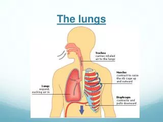
Inspiration • Process of drawing air into lungs (inhalation) • Results from increasing volume of thoracic cavity by inspiratory muscles • Main inspiratory muscles: _________ and ____________ intercostal muscles • External intercostals located in external portion of intercostal spaces (between ribs) • Diaphragm enlarges the thoracic cavity by flattening out.
Expiration • Process of pushing air out of lungs (exhalation) • Results from decrease in size of thoracic cavity • Main expiratory muscles: ___________ intercostal muscles and ____________ muscles • Internal intercostal muscles located between the ribs, deep to the external intercostal muscles • Contraction of abdominal muscles pushes abdominal organs against the diaphragm and pushes diaphragm back into its full dome shape.
Respiratory Volumes • ___________ volume – volume of air inspired and expired during one breath. • Varies according to body’s needs. • Smaller when animal is at rest and larger when excited and active. • __________ volume – volume of air inspired and expired during one minute. • Calculated by multiplying the tidal volume by breaths per minute. • Measured in mL or Liters • __________ volume – volume of air remaining in the lungs after maximum expiration. • Residual volume always remains, lungs will never be completely emptied of air.
Alveolar Gas Exchange- REVIEW!! • Simple _________ of gas molecules from areas of _____ concentration to areas of _____ concentration. • _____ diffuses from the alveolar air into the blood of the alveolar capillary • _____diffuses from the blood into the alveolus
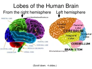
Respiratory Center • Even though all of the inspiratory and expiratory muscles are skeletal muscles under voluntary control, breathing does not require a conscious effort. • Breathing is controlled by an area in the _________ ___________ of the brain stem known as the Respiratory Center. • Directs timing and strength of contraction • Can be consciously controlled for brief periods, therefore the muscles are considered “voluntary”
Mechanical Control System • __________ receptors in the lungs set limits on routine resting inspiration and expiration. • Respiratory center sends out nerve impulses when lungs inflate to a certain point • Stops muscle contractions that produce inspiration and starts contractions to produce expiration • Another set of nerve impulses sent when lungs deflate sufficiently • Stops expiration and starts the process of inspiration again
Chemical Control System • Adjusts normal rhythmic breathing pattern produced by mechanical control system • Chemical (peripheral) receptors in carotid artery and aorta monitor blood _____, ____, and ____. • Central chemical receptors are located in the medulla oblongata.
Chemical Control System • Blood level of CO2 and blood pH are usually linked • __CO2 in blood and __blood pH triggers respiratory center to increase rate and depth of respiration • __CO2 in blood and __blood pH triggers respiratory center to decrease rate and depth of respiration
Chemical Control System • _______ - decrease in blood O2 level • Slight hypoxia triggers respiratory center to increase the rate and depth of breathing • Severe hypoxia - neurons of the respiratory center can become so depressed that adequate nerve impulses cannot be sent to the respiratory muscles • Can cause breathing to decrease or stop completely