SMALL INTESTINE
390 likes | 763 Vues
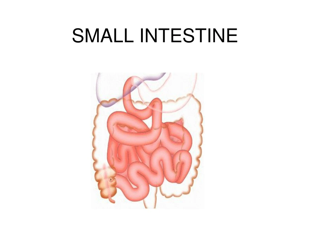
SMALL INTESTINE. SMALL INTESTINE. Terminal part of GIT before it opens into the large intestine. 4 – 6 meters in length. Absorption – main function. Divided into 3 parts:. Duodenum Jejunum Ileum. FEATURES FAVOURING ABSORPTION.

SMALL INTESTINE
E N D
Presentation Transcript
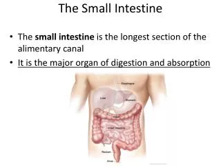
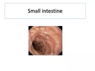
SMALL INTESTINE • Terminal part of GIT before it opens into the large intestine. • 4 – 6 meters in length. • Absorption – main function
Divided into 3 parts: • Duodenum • Jejunum • Ileum
FEATURES FAVOURING ABSORPTION • Extremely long – permits prolonged contact between food and enzymes and also digested products and absorptive cells • Plicae circularis OR Valve of Kerkring – permanent folds of mucous membrane unlike rugae • Villi on plicae circularis • Microvilli on the enterocytes – cytoplasmic extensions
GENERAL HISTOLOGICAL PATTERN OF GIT From inwards out 4 layers are present: • Mucosa • Sub mucosa • Muscularis externa • Serosa OR Adventitia
GENERAL HISTOLOGICAL PATTERN OF GIT • From inwards out 4 layers are present: • Mucosa- consists of : • Lining epithelium – simple columnar cells with striated border. • Layer of connective tissue- lamina propria • Muscularis mucosa- smooth muscles All the above are thrown into folds called ‘villi’ • Sub mucosa- loose areolar tuissue • Muscularis externa – inner circular and outer longitudinal smooth muscles • Serosa OR Adventitia
MUCOSA • Lining epithelium – • Absorptive enterocytes • Goblet cells - mucous secreting • Paneth cells – basally placed exocrine serous cells having zymogen granules. • Argentaffin cells - stimulates smooth muscles by secreting 5 HT. • Crypts of Liberkuhn – invagination of lining epithelium forming simple tubular glands. Basal cells are proliferating cells.
Lamina propria – connective tissue containing blood vessels and lymphoid nodules. • Muscularis mucosa – smooth muscle layer contraction of which results in mixing of food.
DUODENUM • C shaped initial smallest portion of the small intestine • Connects stomach to jejunum • Villi – long, broad, leaf like and numerous • Lamina propria- crypts of Lieberkuhn(STRAIGHT TUBULAR GLANDS) • Sub mucosa – Brunners gland: The identifying feature of duodenum are mucous acini (tubulo alveolar glands) whose duct open into crypts of liberkuhn. The secretion contains bicarbonate and mucous. • Lymphatic nodules are also seen sometimes.
JEJUNUM • Connects duodenum to ileum • Follows the basic histological pattern of GIT • Villi are smaller and less numerous • Goblet cells increase in number • Solitary lymph nodules present
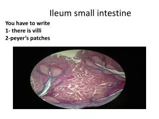
ILEUM • Connects jejunum to large intestine • Fingerlike, smaller and fewer villi • Much more goblet cells seen • Payer’s patches in the sub mucosa also called intestinal tonsils are the identifying features.
INTRINSIC NERVE SUPPLY • Myenteric plexus of Auerbach in muscularis externa. • plexus of Meissner’s – in the sub mucosa.