Download
1 / 9
90 likes | 104 Vues
This study explores the role of caveolae, membrane structures found in various cells, in endocytosis, cell fusion, membrane recycling, lipid rafts, and cell signaling. The loss of caveolae and caveolin-1 gene disruption leads to vascular dysfunction and pulmonary defects in mice.

E N D
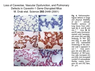
Loss of Caveolae, Vascular Dysfunction, and Pulmonary Defects in Caveolin-1 Gene-Disrupted Mice M. Drab etal. Science 293 2449 (2001)
Biological Membrane Deformation & The aggregation of Membrane proteins The exemple of Caveolae Location Plasma membrane of many cells: Endothelial cells, adypocytes, cardiac muscles… Function Many: Endocytosis - ligand binding Interaction with signaling proteins - cholesterol transport… cell membrane cell interior clathrin-coated vesicle caveolae
“Curvature active” Proteins : Drive Membrane Deformation Endocytosis - Cell fusion Membrane recycling … Ex: endocytosis by formation of Clathrin coated pits Concentrate binding sites Lipid Rafts Cell signalling … Ex. Caveolae Target molecules receptors
Martin Stahlhut, Kirsten Sandvig and Bo van Deurs Experimental Cell Research 261, 111–118 (2000) Hypothetical model of the principal actions of caveolae and caveolins in signaling. Left of dashed line: The major part of caveolins (brown) is present as oligomers in structurally defined caveolae. Filamin (turquoise)– caveolin interactions link some caveolae to actin filaments (tan). Caveolin molecules with a ligand-binding site (scaffolding domain) not involved in oligomer formation can instead sequester and inhibit signaling proteins such as H-Ras (yellow). Activated growth factor receptors (blue-gray) in caveolae recruit adaptor proteins (red-white) like Grb2 and mSOS and can activate caveola-resident H-Ras. Outside of caveolae a fraction of caveolin-1 associates with integrins (gold) and keeps Src-family kinases like Fyn (orange) in an inactive conformation. Upon cell–matrix adhesion (integrin ligation) caveolin-1 and Fyn are coclustered with the integrins, and in the presence of GPI-linked uPAR (red) glycolipid rafts are recruited to the adhesion site. Fyn is activated and the inhibitory action of caveolin-1 is relieved. Fyn signals, via adapter molecules (Shc, Grb2/mSOS), to H-Ras. The activation of H-Ras (in rafts or caveolae) eventually leads to the activation of MAP kinases and signaling to the cell nucleus. K-Ras (light yellow) is present in a different membrane compartment from caveolins due to a polybasic region (pink) and takes part in different signaling events. Raft and caveola membranes are indicated in green. Small vertical lines within the noncaveolar membranes indicate cholesterol concentration. Fyn, mSOS, and Ras associate with the plasma membrane via lipid modifications (black dots). Right of dashed line: Cholesterol depletion of the membrane leads to the loss of the caveolin coat from the membrane. Concomitantly caveolae and functional rafts disappear. This prohibits the local enrichment of H-Ras, Src kinases, adapters, and uPAR. Thus, signaling via caveolin is abolished.
M. Stahlhut etal. Experimental Cell Research261, 111–118 (2000) Epon section of a portion of a rat adipocyte. (BL): basal lamina, (SER): smooth ER (FD): fat droplet Caveolae pictures T. Fujimoto etal. J. Electron Microscopy47, 452 (1998) 100 nm. Electron micrograph of caveolae in the rat smooth muscle cell Barr=100nm As M . Gumbleton Adv. Drug Delivery Rev. 49 (2001) 281 TEM of the alveolar-pulmonary capillary barrier in rat lung (As: Alveolar space; C: capillary lumen) C
N-terminal 1-101 aa hydrophilic attractive part 61-101 aa C-terminal 135-178 a.a Specific attractions N-terminals: aggregation C-terminals: organization hydrophobic transmembrane domain 102-134 Caveolin aggregates 14-16 molecules : 4-6 nm Caveolae Bud 50-80 nm Caveolin - 178 aa Main constituent of Caveolae : Protein Caveolin from Schlegel - Lisanti Cell Signal 10, 457 (1998) 10 nm 100 nm 5 nm
Cf. Surfactant Micelle Caveolae coat structure I. Fernandez, Y. Ying, J. Albanesi & R. Anderson PNAS (2002) 99 pp 11193–11198