Autopsy
Autopsy. An autopsy is a medical procedure that consists of examination performed on a body after death, to evaluate disease or injury that may be present and to determine the cause and manner of a person's death. .

Autopsy
E N D
Presentation Transcript
An autopsy is a medical procedure that consists of examination performed on a body after death, to evaluate disease or injury that may be present and to determine the cause and manner of a person's death.
An autopsy—also known as a post-mortem examination or obduction—is a highly specialized surgical procedure • It is usually performed by a specialized medical doctor called a pathologist.
The number of autopsies performed in hospitals has been decreasing every year. • Reduction in autopsies is negatively affecting the care delivered in hospitals, because when mistakes result in death, they are often not investigated and
Medico-Legal Autopsy or Forensic or coroner's autopsies • Seek to find the cause and manner of death and to identify the decedent. They are generally performed, as prescribed by applicable law, in cases of violent, suspicious or sudden deaths, deaths without medical assistance or during surgical procedures.
Clinical or Pathological autopsies • Are performed to diagnose a particular disease or for research purposes. They aim to determine, clarify, or confirm medical diagnoses that remained unknown or unclear prior to the patient's death.
Anatomical or academic autopsies • Are performed by students of anatomy for study purpose only. • When a person has given permission in advance of their death, autopsies may also be carried out for the purposes of teaching or medical research.
An autopsy is frequently performed in cases of: • Determine as precisely as possible what caused the death. • When a medical condition has not been previously diagnosed. • Doctor is not able to write a death certificate • If there are questions about an unexpected death that appears due to natural causes. • When the death occurs unexpectedly during medical, dental, surgical, or obstetric procedures. • When the cause of death could affect legal matters (when death is believed to result from an unnatural cause). • When the death occurs during experimental treatment
Confirm or exclude a disease diagnosis made before death. An autopsy also may be done to help understand how a given disease progresses or to determine the effectiveness of the treatment for that disease. • Document the presence of a disease that was undiagnosed before death. • Collect samples of body fluids or tissues for possible genetic testing. This is generally done only after discussion with the deceased person's family. • Collect evidence and information in criminal cases. • Help health departments or other government agencies identify and track a disease or potential public health hazard (such as a suspected contagious disease or contaminated drinking water).
Before the actual autopsy, as much information as possible is gathered about the person who died and the events that led to the death. This includes reviewing medical records and consulting with the person's doctors about previous medical problems.
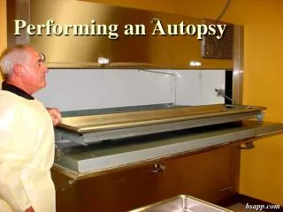
The body is opened using a Y-shaped incision from shoulders to mid-chest and down to the pubic region. If the head is to be opened, the second incision is across the head, joining the bony prominences just below and behind the ears. • The autopsy technician uses a scalpel or a special knife for the incisions. There is almost no bleeding, since a dead body has no blood pressure except that produced by gravity. • The organs are taken off on the table in blocks- thoracic organs, abdominal organs, pelvic organs, brain and they are dissected and grossly examined. Sections for the histopathology are taken if necessary.
Completion of the autopsy may require examination of tissues under a microscope. • A written report describes the autopsy findings. This report may address the cause of death and may help answer any questions from the deceased person's doctor and family.
After performing the autopsy, the pathologist will generally make a statement about the cause of death. Some examples of natural causes include: • Damage to the heart caused by heart disease, a heart attack, or heart failure. • Damage to the brain caused by conditions such as tumors, bleeding, stroke, poorly controlled epilepsy, diabetes, or Alzheimer's disease. • Damage to the lungs caused by a blood clot, bleeding, or pneumonia. • Damage to organs in the abdomen, such as the stomach, spleen, liver, or kidneys.
An unnatural death means the death resulted from an unexpected, unusual, or suspicious cause. If an injury caused or contributed to the death, the manner of death is called unnatural. Unnatural manners of death are homicide, suicide, accident, and undetermined. Unnatural deaths generally are investigated under authority of the medical examiner or coroner, and the determination of the manner of death requires a detailed investigation of the circumstances surrounding the death. Some unnatural causes of death include: Bullet wounds. An automobile accident or plane crash. Fire, drowning, or electrocution. Death resulting from extreme heat or cold. Poisoning or drug overdose.
The autopsy begins with a careful examination of the external part of the body. Photographs may be taken of the entire body and of specific body parts. The location and description of identifying marks, such as scars, tattoos, birthmarks, and other significant findings (injuries, wounds, bruises, cuts), are recorded on a body diagram.
Gross examination and description of organs and tissue samples
Position and shape of organ • Comparison with the „normal” shape or organ form (e.g. deformed, not deformed -„normal”)
Size and Weight • Both quantities are determined by comparison with normal organ size and weight (e.g. decreased, increased) • Size - length, width and height - always given in centimeters • Weight - always given in gram (the most reliable measurement of the organ size)
Color • Blood content: • bright red (active hyperaemia); • dark blue-red (dusky) - congestion (passive congestion); • black-red (haemorrhagic infarction); • Pigments: • yellow/ red-yellow (bilirubin); • black (melanin-naevi, • malignant melanoma, • carbon- anthracosis); • brown (hemosiderin, lipofuscin)
Yellow: fat; • intensive yellow (e.g. adrenal cortex, corpus luteum accumulation of steroids), • Necrotic tissue: • pale (e.g. fresh infarction of the cortex of the brain); • black-red (haemorrhagic infarction); • yellow/ gray- yellow (anaemic infarction e.g. heart) • Others: • gray-white (e.g. neoplastic tumours with little vascular supply; • large fibrin deposits); • white (cholesterol deposits e.g. atherosclerosis; fibrotic scar);
Consistency (determined by touch) • Firm- e.g. the liver in cirrhosis; the lung in silicosis, • Hard- calcium or bone, • Medium-firm- certain carcinomas , • Doughy- fatty liver, • Soft diffluent- septic spleen, • Acute liver atrophy, • Fragile breakable - foci of pneumonia, gangrene of the lung, • Elasticity- some normal organ and tissue
External Surface • Smooth – uneven – irregular (scars) • Shiny- mat, dull (fibrin exudate) • Dry • Smooth- granular- nodular- lobular- folded • Cut surface • (like external surface) • transparency (e.g. nodular goiter)
Findings (Focal Finding, Lesion) • Localization • Topography • Size (lenght, width and height – in centimeters) • Form: round, oval, irregular shape • Type of delineation • Sharp • Indistinct • with (fibrous) capsule/ encapsulated • Color • Consistency
External Inspection • Remove any bandages and document any therapeutic devices • Remove superficial, peripheral venous catheters, but leave indwelling central lines, endotracheal tubes, feeding tubes, urinary bladder catheters in place until the internal examination confirms their proper location
Measure the body length, and if possible, weight it • Identify any abdominal distention and determine whether the extremities are symmetrical • Document abnormalities by measuring the circumferences of the chest (at level of the nipples), abdomen (at the umbilicus) or extremities
Inspect the skin anteriorly and posteriorly • Note its color and elasticity and characterize any cutaneous lesions • Document surgical and nonsurgical scars • Record any tattoos or other identifying features • Examine the character and colour of the nails
Estimate the degree of rigor mortis by flexing the joint • Inspect and palpate the skull and breast • Examine the eyes