Download
1 / 49
2.08k likes | 7.2k Vues
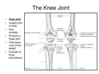
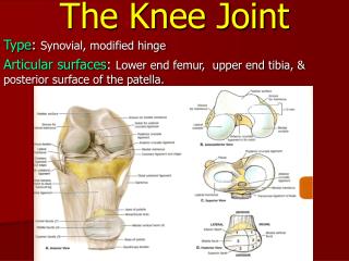
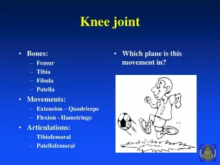
Regional Biomechanics knee Joint. Kinematics Kinetics Pathomechanics. Biomechanics of the knee joint 1- Bony Structure. 1- Femoral articulation: pulley-shaped, convex, longer anteroposteriorly than transversely. - Shaft of the femur is not vertical.

E N D
Regional Biomechanicsknee Joint Kinematics Kinetics Pathomechanics
Biomechanics of the knee joint1- Bony Structure 1- Femoral articulation: pulley-shaped, convex, longer anteroposteriorly than transversely. - Shaft of the femur is not vertical. - Medial condyle 2/3 of an inch longer than the lateral 2- Tibial Articulation: Concave -medial tibial condyle is 50% larger than the lateral condyle * All articular surfaces are covered by cartilage with a thickness of 3-4mm.
Tibiofemoral Angle • The mechanical axis of LE (WB line) passes from the center of the femur to the superior surface of head of talus (average 3 deg. with the vertical). • Frontal plane. Measured between anatomical axes of femur and tibia. • Normal value 170-175 laterally or 185-190 medially. • Laterally: less than 170 “Genu valgum or Knock-knee” • more than 180 “Genu varum or bow-leg”
(A) Genu valgum (b) Genu varum • (A) (B)
The Q (quadriceps) angle • Frontal view. Between a line connecting between the ASIS to the mid point of patella and a line connecting the tibial tubercle to the mid point of patella. • Normal value 15 deg. • Greater in females than males due to wider pelvis and increases femoral anteversion.
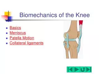
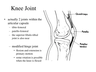
2- Capsule of the Knee joint • Reinforced - Posterior: by muscles (popliteus, gastrocnemius, and hamstring) ligaments (oblique popliteal, and arcuate popliteal ligament). • Lateral & Medial: medial and lateral patellar retinacular fibers, collateral ligaments and iliotibial band. • Anterior: Quadriceps tendon, patella and patellar ligament, .
3- Menisci of the knee jointFibro cartilaginous joint discs Attach to intercondylar region of the tibia
Medial meniscus • C- shaped and is attached to the medial collateral ligament and to the semimembranosus muscle. It is more firmly attached and less movable so it is more torn than the lateral meniscus. • Lateral meniscus • 4/5 of a ring, is much loose and mobile than the medial meniscus • The ante anterior horns of the two menisci are linked by the transverse ligament.
Function Medial meniscus & Lateral meniscus1- Distribute weight.2- Increase the joint congruency.3- Lubricate the articular cartilage.4- Reduce friction between joint surface.5- Shock absorber.
4- Ligaments of the knee joint(1) Medial collateral ligament • Position: Medial aspect of the joint. • Attachment: Med. Femoral epicondyle and upper end of tibia. • Orientation: Inferior & Anterior. • Function: 1- Resist valgus stress especially when knee is extended. 2- Resist lateral rotation of tibia. 3- Restrict Ant. Displacement of tibia. 4- Resist Excessive Knee extension.
(2) Lateral collateral ligament • Position: Lateral aspect. • Attachment: Lat. Epicondyle. Head of fibula. • Orientation: Inferior and Posterior. • Function: 1- Resist varus stress. 2- Resist axial rotation. 3- Resist Post. Displacement of tibia. 4- Resist knee extension. N.B: Both collateral ligaments are relaxed at 20-30 flexion so it is the position of immobilization after injury.
(3) Posterior Capsular ligament 1)Oblique popliteal ligament. • Position: Posteromedial aspect. • Attachment: Med. Tibial condyle central part of posterior aspect of the joint capsule. • Orientation: Upward and laterally. • Function: 1-Check valgus stress. 2-Tight in full extension. 2)Arcuate popliteal ligament. • Position: Posterolateral aspect. • Attachment: Post, aspect of the head of fibula lat. epicondyle • Orientation: upward and medially. • Function: 1- Check varus stress. 2- Tight in full extension.
4- Anterior Cruciate Ligament • Position: Intracapsular ligament. • Attachment: Ant. Part of intercondylar eminence post part of inner aspect of lat. Femoral condyle. • Orientation: Posterior, Superior and lateral. • Function: 1- Prevent anterior displacement of tibia 85%. 2- Limit full knee extension. 3- Resist varus and valgus stresses (minor contribution). 4- Control medial rotation (axial) of the tibia. N.B: Injury to the ACL occurs when the knee is flexed and the tibia rotates in either direction.
5- Posterior Cruciate Ligament • Position: Intracapsular Ligament. • Attachment: Post. Part of intercondylar eminence Anterior part of inner aspect of medial femoral condyle. • Orientation: Ant. superior, and medially. • Function: • Prevent posterior displacement of tibia 95%. • Tight during full flexion. • Resist varus and valgus stresses (minor contribution).
Function of cruciate ligaments during knee motion. • Full extension: ACL is more vertical & PCL is more horizontal. • During hyperextension ACL is stretched and PCL is relaxed. • Full flexion: PCL raised up vertically making 60 degrees with tibia and become taut. • Medial rotation: ACL wind around PCL ( ACL stretches and PCL relaxes). • Lateral rotation: parallel “ACL relax and PCL stretches”
6- iliotibial band • Position: Anterolateral aspect of the knee joint. • Attachment: fascia of tensor fascia lata, G. max, and G. med, lateral tubercle of tibia. • Orientation: two band one downward and the other Anterior and lateral to patella “Iliopatellar band”. • Function: 1- Tight regardless the position of the hip or the knee. 2- Prevent post. Displacement of femur when the tibia is fixed and knee extended.
Stability of the knee joint • Stability of the knee joint is provided by: -Static stabilizers (joint capsule and powerful ligaments) • - Dynamic stabilizers (flexor and extensors muscles)
Stability of the knee joint • Close-packed position: Max Stability Max. Extension and max. lateral rotation. “Screw home mechanism” • Loose-packed position: Min. Stability Flexion position
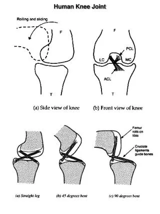
Knee axis of motion • The axis of knee motion passes horizontal and oblique through the knee joint (lower on the medial side). So full flexion is accompanied by medial tibial rotation and full extension is accompanied by lateral tibial rotation . • This axis moves through the ROM forming a semicircle moving posteriorly and superiorly on the femoral condyles with increasing flexion (instantaneous axis of rotation -IRA).
Surface motion of the knee joint during flexion in CKC During flexion: - From full extension to 25º of flexion is pure posterior rolling of the femoral condyles on the tibia. - After 25º rolling is accompanied by anterior gliding to prevent posterior dislocation of the femoral condyles (facilitated by the ACL). - At the end of the range of flexion the femoral condyles glide without rolling
Surface motion of the knee joint during extension in CKC • During extension: • The first part of the extension range is pure anterior rolling of the femoral condyles on the tibia displacing them back to the neutral position. • After that anterior rolling is accompanied with posterior gliding ( facilitated by the PCL).
Role of the menisci during flexion and extension • During flexion : the menisci move posteriorly: the MM moves posteriorly by the semimembranosus while the LM is drawn posteriorly by the popliteus • During extension : the menisci are pulled anteriorly by the meniscopatellar fibers. The posterior horn of the LM is pulled anteriorly by tension in the meniscofemoral ligament.
Role of Cruciate ligament • During flexion: ACL causes the femoral condyle to slide ant. while the femur rolls posteriorly. • During extension: PCL causes the femoral condyle to slide post. While the femur rolls anteriorly.
Load transmission through the knee joint • Distal end of femur: • (a)-Vertical lateral trabeculae Ipsi(compression force). Contralateral (tension force). • (b)- Horizontal trabeculae : join the two condyle. • Distal end of tibia: the similar set
Load transmission through the intact menisci • Collagen in the menisci are oriented in circumferential direction. • Load on the knee joint will cause extruding force(which pushes the menisci outward) • This force is resisted by their powerful attachment to the tibia. • In their resistance the menisci transmit some of the load to the tibia.
Load transmission through torn menisci • The meniscus become redundant. • During transmission of load , the meniscus will not be able to resist the extruding forces so it will open and loads will be transmitted directly between condyles. • The load will be carried by the cartilage . This will increase the joint load significantly.
Patellofemoral Jointsurface motion of the patella on the femur • During flexion: • The patella slides distally between the femoral condyles (travels twice its length(8cm). • The patella moves also backward or posteriorly. • - Tilts medially (rotate around its vertical axis).11° “Vertical axis”(from 25 to130 flexion) • The patella also rotate medially around its Anteroposterior axis (medial rotation). • SO(during flexion: the patella slides downward and moves posteriorly and medially. In addition it tilt and rotate medially.
Patellofemoral Jointsurface motion of the patella on the femur • During extension: the patella slides upward and moves anteriorly away from the femoral condyles and laterally. In addition it tilt and rotate laterally.
Function of the patella • Improve mechanical efficiency of the quadriceps muscle through two mechanism: 1- Increase the moment arm. 2- Increase the angle of pull. • Reduce friction between the quadriceps tendon and femoral condyles. • Provide good cosmetic appearance.
The mechanical effect of patella on the moment arm through the ROM in addition to the physiological effect • In full knee flexion: the patella moves downward and backward on the intercondylar groove. So the moment arm of the quadriceps decreases. This does not affect the torque because of two reasons. (1) – the IAR moves posteriorly away from the line of action of the quadriceps. (2) – The muscle at physiological advantage as it is stretched (length- tension relationship).
The mechanical effect of patella on the moment arm through the ROM in addition to the physiological effect • During knee extension: The patella moves upward and forward on the intercondylar groove. So the MA of the quadriceps lengthens ( mechanical advantage). The maximum torque of the quadriceps is produced at 60º because the muscle shows both mechanical and physiological advantaged. • With continuous extension: the MA again begins to diminish.
The mechanical effect of patella on the moment arm through the ROM in addition to the physiological effect • At the last 15º of extension: the quadriceps at both mechanical disadvantage (decrease MA) and physiological disadvantage (decrease muscle length). A 60% increase in force is required to complete the range.
Effect of removal of the patella: • Removal of the patella decrease the quadriceps torque up to 50%. • N.B: Loss of patella has its most apparent effect in the last stages of extension when there is both mechanical and physiological disadvantages especially if the muscle has to work against the resistance of gravity.
Stability of the Patellofemoral joint(mediolateral forces on the patella) • During full extension and the quadriceps is relaxed: the patella can be passively displaced medially or laterally half the width of the patella (so it is used as position for patellar mobilization). • During active extension: the force of the patella is determined by the pull of the quadriceps and the patellar tendon. Since they do not lie in the same action lines, the patella tends to be pulled laterally. This may cause the patella to sublaxate or dislocate laterally.
Mediolateral forces on the patella • The patellar tendency towards lateral dislocation is prevented by: • (1)- the lateral lip of the patellar surface of the intercondylar groove. • (2)- The muscular pull of vastus medialis longus and vastus medialis oblique (VMO) muscles.
Risk factors for lateral patellar dislocation Tightness of iliotibial band Laxity of MCL Increased Q angle Increased genu valgum Excessive hip anteversion Excessive external tibial torsion Weakness of VMO Shallow patellar track
Kinetics ( Patellofemoral JRF) • During full flexion : the patella sinks and becomes more in contact in the intercondylar groove. So the compressive forces (JRF) increases. • Between 90º- 70º of knee flexion: the quadriceps tendon contacts the femoral condyles and dissipates some of the PF compression. • During full extension: the patella makes little or no contact with the femur so the compressive forces decreases. That is why straight leg raising is used to improve the quadriceps strength in cases of PF problems.
Patellofemoral joint reaction force values JRF is ½ W at 15 knee flexion. JRF 7.8W at 130 knee flexion. JRF 3.3 W during climbing stairs.
Calculation of PF joint reaction force • R= 2T cos Ø/2
Pathomechanics of the knee joint1- Bony abnormality: Genu varum (bow legs): Tibiofemoral less than normal medially. Center of joint displaces laterally leading to medial osteoarthritis.
Genu Valgum “Knock knees”: Tibiofemoral greater than normal medially. Center of joint displaces medially leading to lateral osteoarthritis.
2-Meniscus injury(A) twisting movement of the knee(B) violent extension of the knee
3- Ligaments injury1) anterior cruciate ligament • Mechanism of injury: foot firmly planted and femur vigorously externally rotated or translated posteriorly. Another mechanism excessive hyper extension of the knee. • Following injury: hamstring spasm. • Post surgical rehabilitation: • Exercise for hamstring and quadriceps to keep the ratio of 0.7 : 1 • Avoid OKC exercise for the first 3 months. • CKC exercises are the choice for early post operative rehabilitation.
2) posterior cruciate ligament • Mechanism of injury: 1- Falling over hyper flexed knee. 2- dash board injury. • Rehabilitation program directed for strengthening quadriceps to prevent posterior displacement of tibia
4- Patellar dysfunction1) Change of Q angle2) Chondromalachia patella 3) Patellectomy: - Reduce the torque of quadriceps by about 49% - Internal moment arm of quadriceps reduce from 4.7 cm to 3.8 cm. - Has no effect on the strength of quadriceps if the knee fully flexed. - Has effect at the last stage of knee extension.