Detection and Characterization of DrsG in Streptococcus dysgalactiae subsp. equisimilis
This study presents evidence for the presence of the gene drsG in Streptococcus dysgalactiae subsp. equisimilis (SDSE) using PCR and Southern blot analysis, showcasing various isolates and their associated emm-types. The structural features of DrsG proteins were examined through schematic diagrams and Western blot assays to determine their expression and activity. Notably, DrsG was found not to inhibit complement-mediated lysis of erythrocytes in certain assays. The interaction of LL-37 with DrsG proteins was also investigated, revealing insights into their potential role in bacterial survival.

Detection and Characterization of DrsG in Streptococcus dysgalactiae subsp. equisimilis
E N D
Presentation Transcript
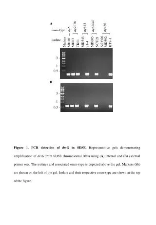
stg62647 stg2078 A stg6 stg643 stg480 emm-type NS3396 MD592 MD543 MD605 Marker TK01 NCU21 KTS-1 81-4 isolate MD10 MD03 3 - 1 - 0.5 - B 3 - 1 - 0.5 - Figure 1. PCR detection of drsG in SDSE. Representative gels demonstrating amplification of drsG from SDSE chromosomal DNA using (A) internal and (B) external primer sets. The isolates and associated emm-type is depicted above the gel. Markers (kb) are shown on the left of the gel. Isolate and their respective emm-type are shown at the top of the figure.
emm65 stg93464 stg2078 stg2078 emm12 stg6 emm1 emm-type Marker MD128 M12 ES61 MD10 MD03 M1 TK01 Isolate 6.6 - 4.4 - 2.3 - Figure 2. Chromosomal detection of drsG in SDSE. Representative southern analysis of KpnI digested SDSE chromosomal DNA probed with a DIG dUTP labeled internal 250bp fragment of drsG. Molecular weight markers are shown on left.
0 50 100 150 200 250 300 RD2 RD1 SS DrsGL DrsGS C-term SRR DRS PRR C-term PRR SRR LRR SIC Figure 3. Schematic diagram showing the conserved repeat domain structure of DrsSGL, DrsGS, DRS and SIC. For DrGLs and DrsGS, the prolin rich sequences are found within RD2. Abbreviations:Signal Sequence (SS), Repeat Domain 1 (RD1), Repeat Domain 2 (RD2), Short Repeat Region (SRR), Proline Rich Region (PRR), Long Repeat Region (LRR) and C-terminal region (C-term).
A 1 2 3 4 5 36- 25- 19- B 1 2 3 4 5 36- 25- 19- Figure 4. DrsG is expressed and secreted by SDSE. (A) Western blot of concentrated culture supernatants from mid-log and stationary cultures of drsGL-positive GGS124 (lanes 1 and 2), drsG-negative MD985 (lanes 3 and 4) and DrsGL (lane 5) probed with anti-DrsGL antibodies. (B)Western blot of concentrated culture supernatants from mid-log and stationary cultures of drsGS-positive MD604 (lanes 1and 2), drsG-negative MD128 (lanes 2and 3) and DrsGS(lane 5) probed with anti-DrsGSantibodies. Molecular weight markers indicated on left.
Figure 5. DrsG does not inhibit complement mediates lysis of erythrocytes.DrsGL, DrsGS, SIC or PBS was pre-incubated with human serum. Sensitised sheep erythrocytes were then added and incubation continued for a further 30min. Unlysed erythrocytes were removed by centrifugation and haemolysismeasured by measuring the absorbance of the supernatant at 415nm. Results are presented as inhibition of complement mediated lysis, whereincubation of sheep erythrocytes with water was used the reference point for 100% lysis. Data represent the mean of three independent experiments. Statistical significance was determined using the unpaired t-test.
A B C Figure 6. LL-37 interaction with DrsGL, DrsGS and SIC. LL-37 interactions with DrsGL, DrsGS and SIC. Graphs on the left of each panel show the results when DrsGL, DrsGS or SIC were coated to the wells of a 96-well plate. LL-37 was added, followed by anti-LL-37 antibody, HRP-labelled secondary antibody and horse radish peroxidase. Results for assays in which LL-37 (C1), LL-37 and primary antibody (C2) or secondary antibody (C3) were not added are included as controls. The graphs on the right of each panel represent the reciprocal assay in which LL-37 was used as bound substrate, and free DrsGL, DrsG or SIC added
A B C Figure 7. Growth of SDSE in the presence of LL-37 and DrsG.SDSE MD985 (drsG-negative) was grown in the presence of LL-37 and/or DrsG/SIC. After a 2.5hr incubation, the bacteria were recovered, plated onto Todd-Hewitt agar and incubated overnight. The percentage growth, when compared to controls was determined for each individual assay. The results presented are the mean of three independent experiments.