AIM Project Update: Advances in Medical Imaging Annotation and Markup Technologies
The AIM Project Update presented by Daniel Rubin, MD, and his colleagues outlines significant advancements in medical imaging annotation and markup tools. Highlighting a case study involving a potentially malignant mass in the left upper lung, the presentation emphasizes the role of the AIM 1.0 model in integrating various data formats (XML, DICOM) and standards (SILVER COMPLIANT). The research facilitates improved communication and collaboration in translational biomedical informatics, which is essential for enhancing patient care and facilitating research.


AIM Project Update: Advances in Medical Imaging Annotation and Markup Technologies
E N D
Presentation Transcript
AIM ProjectUpdate March, 2009
Presenters Daniel Rubin M.D., M.S. Assistant Professor of Radiology Research Scientist, Center for Biomedical Informatics Research Stanford University David S. Channin, MD Associate Professor of Radiology Northwestern University Pattanasak Mongkolwat PhD Research Associate Professor of Radiology Northwestern University Vladimir Kleper Member of the Technical Staff Northwestern University
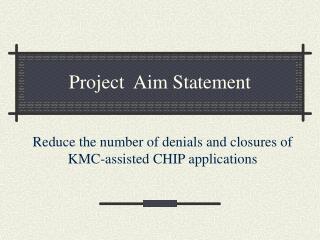
What’s the problem? “There is a spiculated, somewhat microlobulated, approximately 2 cm mass of soft tissue attenuation in the left upper lobe suspicious for malignancy”
An Image, A Markup, An Annotation Anatomic Entity: Left Lung (Radlex:1326) Anatomic Entity: Upper lobe of left lung (RID1327 Observation: Mass (RID:3874) Characteristic: Microlobulated margin (RID5712) Geometric Shape: Polyline 2D coordinates: {(x,y),(x,y)….} Calculation: Largest diameter result: 2.8 cm
AIM Update • AIM contracted ended 9/30/08 • AIM 1.0Rev12 is SILVER COMPLIANT! • AIM UML Model • AIM XML Schemas • AIM SDK to create AIM Instances • In XML, DICOM, HL7 CDA • ANIVATR • Open Source reference implementation to validate and translate AIM instances • ALL Available at cabig.nci.nih.gov/tools/AIM • Publications • Rubin D, Mongkolwat P, Kleper V, Channin DS. Medical imaging on the semantic web. The Association for the Advancement of Artificial Intelligence. (AAAI) Spring Symposium, Palo Alto, CA, 2008. • Channin DS, Mongkolwat P, Kleper V, Sepukar K, Rubin, DL. The caBIG Annotation and Image markup Project. J Digit Imaging. 2009 (in press). • Rubin DL, Mongkolwat P, Channin DS. A Semantic Image Annotation Model to Enable Integrative Translational Research. 2009 AMIA Summit on Translational Bioinformatics. San Francisco. 2009. • Channin DS, Mongkolwat P, Rubin DL. Editorial: Small Project, Big AIM. Radiology (in revision).
What the AIM 2.0 UML Model would look like if AIM 2.0 existed
Annotation Image Annotation Annotation of Annotation User Equipment Patient Image Reference DICOM Images Web Images Anatomic Entity Anatomic Entity Characteristic (2.0) Rating (2.0) Imaging Observation Imaging Observation Characteristic Rating (2.0) Inference (2.0) Probability Map (Segmentation Map(2.0)) Text Annotation Geometric Shape 2D and 2D coordinates Multipoint, point, circle, ellipse, poly Calculation Calculation Result Included in the model
Rating class • max (Double) • min (Double) • name (String) • value (Double)
Segmentation class • type (Value Domain) • Binary • Fractional Probability • Fractional Occupancy • Surface • instanceUID (String) • sopClassUID (String) • referencedInstanceUID (String) • segmentNumber (Integer)
Inference class • codeMeaning (String) • codeSchemeDesignator (String) • codeValue (String) • codingSchemeVersion (String, option) • confidence (Double, option) • truth (Boolean, option)
ReferencedGeometricShape • referencedShapeIdentifier (Integer)