Acute Coronary Syndromes
370 likes | 683 Vues
Acute Coronary Syndromes. Acute Coronary Syndrome. Definition : a constellation of symptoms related to obstruction of coronary arteries with chest pain being the most common symptom in addition to nausea, vomiting, diaphoresis etc.

Acute Coronary Syndromes
E N D
Presentation Transcript
Acute Coronary Syndrome Definition: a constellation of symptoms related to obstruction of coronary arteries with chest pain being the most common symptom in addition to nausea, vomiting, diaphoresis etc. Chest pain concerned for ACS is often radiating to the left arm or angle of the jaw, pressure-like in character, and associated with nausea and sweating. Chest pain is often categorized into typical and atypical angina.
Typical anginal pain —All three of the following • Substernal chest discomfort • Onset with exertion or emotional stress • Relief with rest or nitroglycerin • Atypical anginal pain • 2 of the above criteria • Noncardiac chest pain • 1 of the above
Acute coronary syndrome • Based on ECG and cardiac enzymes, ACS is classified into: • STEMI: ST elevation, elevated cardiac enzymes • NSTEMI: ST depression, T-wave inversion, elevated cardiac enzymes • Unstable Angina: Non specific EKG changes, normal cardiac enzymes
Unstable Angina Occurs at rest and prolonged, usually lasting >20 minutes New onset angina that limits activity Increasing angina: Pain that occurs more frequently, lasts longer periods or is increasingly limiting the patients activity
EKG • STEMI: • Q waves , ST elevations, hyper acute T waves; followed by T wave inversions. • Clinically significant ST segment elevations: • > than 1 mm (0.1 mV) in at least two anatomical contiguous leads • or 2 mm (0.2 mV) in two contiguous precordial leads(V2 and V3)
EKG • NSTEMI: • ST depressions (0.5 mm at least) or T wave inversions ( 1.0 mm at least) without Q waves in 2 contiguous leads with prominent R wave or R/S ratio >1. • Isolated T wave inversions: • can correlate with increased risk for MI • may represent Wellen’s syndrome: • critical LAD stenosis • >2mm inversions in anterior precordial leads
Cardiac Enzymes • Troponin is primarily used for diagnosing MI because it has good sensitivity and specificity. • CK-MB is more useful in certain situations such as post reperfusion MI or if troponin test is not available • Other conditions can cause elevation in troponin such as renal failure or heart failure • The increasing troponin trend is the important thing to look for in diagnosing MI. Order Troponin together with ECG when doing serial testing to rule out ACS.
Pathophysiology • ACSs result from myocardial ischemia due to imbalance between myocardial O2 demand & supply • usually due to occluded coronary artery thrombus • STEMI, NSTEMI, & unstable angina fall under this heading • unstable angina does not produce detectable biochemical marker levels
Plaque Rupture & Clot Formation • > 90% of ACSs caused by rupture or erosion of an atherosclerotic plaque • Plaques likely to rupture • those that occlude < 50% of the lumen • eccentric shape • thin fibrous cap with large fatty core • Clots form on top of ruptured plaques & partially or fully occlude the artery lumen • Exposure of collagen & tissue factor from the plaque induces platelet adhesion & activation • promotes release of vasoactive substances
Atheroma Production A: normal muscular artery. The adventitia, or outermost layer of the artery, consists principally of recognizable fibroblasts intermixed with smooth muscle cells loosely arranged between bundles of collagen and surrounded by proteoglycans. It is usually separated from the media by a discontinuous sheet of elastic tissue, the external elastic lamina. B: platelet aggregates, or microthrombi, form as a result of adherence of the platelets to the exposed subendothelial connective tissue. Platelets that adhere to the connective tissue release granules whose constituents may gain entry into the arterial wall. Platelet factors thus interact with plasma constituents in the artery wall and may stimulate events shown in the next illustration C: smooth muscle cells migrate from the media into the intima and actively multiply within the intima. Endothelial cells regenerate in an attempt to re-cover the exposed intima, which thickens rapidly owing to smooth muscle proliferation and formation of new connective tissue.
Plaque Rupture & Clot Formation • Platelet activation: changes on platelet GP IIb/IIIa receptors lead to formation of fibrin bridges • inclusion of many platelets creates “white” clots • more common in NTSE ACS • incomplete artery occlusion • Coagulation cascade activated: fibrin traps RBCs • clots have red appearance • more common in STEMI ACS • more likely to completely occlude vessels • Myocardial ischemia can result from microthrombiembolization & lead to necrosis
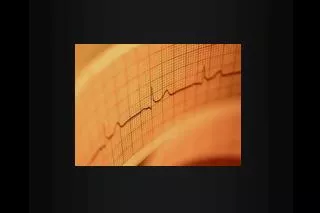
Electrocardiogram • 12-lead ECG should be done within 10 min of ED arrival • Key findings indicating myocardial damage • ST-segment elevation • ST-segment depression • T-wave inversion
Biochemical Markers • Evaluate troponin & CK MB to confirm MI • released in response to myocardial necrosis • 3 measurements taken over the 1st 12 to 24 hrs • MI diagnosis: • > 1 one troponin value greater than MI decision limit set by lab or • 2 CK MB values greater than MI decision limit set by lab
The Three I’s • Ischemia=ST depression or T-wave inversion Represents lack of oxygen to myocardial tissue
The Three I’s • Injury = ST elevation -- represents prolonged ischemia; significant when > 1 mm above the baseline of the segment in two or more leads
The Three I’s • Infarct = Q wave— represented by first negative deflection after P wave; must be pathological to indicate MI
What part of the heart is affected ? • II, III, aVF = Inferior Wall I II III aVR aVL aVF V1 V2 V3 V4 V5 V6
Based on the EKG, which vessel in the heart is blocked? • II, III & aVF = Inferior Wall MI = Right Coronary Artery blockage
I II III aVR aVL aVF V1 V2 V3 V4 V5 V6 Which part of the heart is affected ? • Leads V1, V2, V3, and V4 = • Anterior Wall MI
Based on the EKG, which vessel in the heart is blocked? • V1 - V4 = Anterior Wall (Left Ventricle) = Left Anterior Descending Artery Blockage
What part of the heart is affected ? • I, aVL, V5 and V6 Lateral wall of left ventricle I II III aVR aVL aVF V1 V2 V3 V4 V5 V6
Based on the EKG, which vessel in the heart is blocked? • I, aVL, V5 + V6 = Lateral Wall = Circumflex Artery Blockage