Pathway (Iron Deficiency)
This pathway outlines the management of Iron Deficiency Anemia (IDA) established by the British Society of Gastroenterology in May 2005. It encourages general practices within SWCCG to identify patients with iron deficiency and monitor their care to reduce unnecessary referrals to secondary care. Iron deficiency is confirmed through specific hemoglobin levels and laboratory tests. The guidelines also stress the importance of thorough patient history, dietary assessment, examination, and necessary investigations, especially for high-risk groups, to effectively address and manage IDA.

Pathway (Iron Deficiency)
E N D
Presentation Transcript
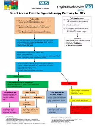
Pathway (Iron Deficiency) Iron Deficiency Anaemia (IDA) Guidelines for QP British Society of Gastroenterology produced the guidelines for the management of iron deficiency anaemia in May 2005. (A F Goddard, MW James, A S McIntyre and BB Scott on behalf of the BSG). This pathway is to encourage and monitor the use of these pathways in general practice in SWCCG to reduce inappropriate referrals to secondary care. Definitions: Anaemia is a haemoglobin below 13 g/dL (130 g/L) in men over 15 yrs, below 12 g/dL (120 g/L) in non pregnant women over 15 yrs, and below 11 g/dL (110 g/L) in pregnant women If there is anaemia, iron deficiency should be confirmed by either a low serum ferritin, (< 15 mg/L) (but <50 mg/L is compatible with iron deficiency) a red cell microcytosis or a hypochromia (in the absence of chronic disease or haemoglobinopathy) From the start date of this QP pathway, practices should identify new patients with markers for iron deficiency (any of the Read codes) and for these patients take a full history and perform an appropriate examination: History: Diet; Medication (NSAIDs); Family History (may indicate inherited malabsorbtion or haemoglobinopathies such as thalassaemia); telangectasia; blood donation. Examination: Urine testing (1% of pts with IDA will have renal tract malignancy, and 1/3 of patients with renal cell carcinoma have IDA). Abdominal exam – pr can be postponed until colonoscopy Other tests: Coeliac screen in all cases, Consider upper and lower GI investigations in all men with iron deficiency AND anaemia, Consider upper and lower GI investigations in women post menopausal or who are over 50 yrs old who have iron deficiency AND anaemia, or who have a strong FH of colorectal cancer. (IDA occurs in 15-20% of healthy premenopausal women and colonic investigation should be reserved in premenopausal women for those with a strong FH of bowel cancer, those with colonic symptoms or persistent IDA following iron supplementation and correction of other potential causes, egmenorrhagia/blood doning/poor diet). (Oesophagogastroduodenoscopy (OGD) should be considered for pre-menopausal women with IDA and upper GI symptoms according to DoH guidelines for suspected upper GI cancer).
Reduce Emergency AdmissionsMDT Meetings and review of those who have died For patients who died since the previous meeting: Were they on the EoL register? – and if not, why not? How complete was the EoL care plan? Did they die at their preferred choice of place of death? What went well? What did not go so well? Are there any bereavement issues? Could/should anything have been done differently?
Reduce A&E Attendances(asthma review) www.asthma.com/resources/asthma-control-test.html
Reduce A&E Attendancesnew D-Dimer pathway Clinically Suspected DVT All patients with a DVT should have a full clinical history and examination undertaken with the aim of detecting an underlying condition contributing to the development of thrombosis. If patients with a suspected DVT fall into any of the following groups, they should be admitted as an emergency to hospital. They should NOT be managed in primary care: • Severe DVT: features include • groin pain • significant colour change in whole of affected limb • involvement of whole leg • Suspected pulmonary embolism • Pregnant • At risk of bleeding: • Active peptic ulcer • Uncontrolled hypertension (>200/>110 mmHg) • Eye or CNS surgery in the last 1 month • CVA in the last 1 month • Known thrombocytopenia (platelets <80)
Reduce A&E Attendances (new D-Dimer pathway) 2-Level DVT Wells Score 2-level DVT Wells score ≥2 requires a scan and a D-Dimer test. They require either LMWH or rivaroxaban whilst awaiting the scan. Where the D-Dimer is positive but the scan negative – they require a repeat scan 6-8 days later. 2-level DVT Wells score ≤ 1 requires a scan ONLY if the D-Dimer is positive. If a diagnosis of DVT is confirmed, then the patient should commence warfarin, or if suitable, continue rivaroxaban