Download
1 / 50
550 likes | 597 Vues
This resource provides insights into benign liver pathologies, including pyogenic abscess, amoebic abscess, hydatid cysts, simple cysts, and polycystic liver disease. It covers causes, clinical features, diagnostic approaches, and treatment modalities for each condition.

E N D
DR. A .PERVEZ (PG, DEPT OF GENERAL SURGERY) BENIGN PATHOLOGIES OF THE LIVER
INTRODUCTION *Benign liver pathologies are broadly divided into two categories *Firstly, infectious diseases such as 1.Pyogenic abscess 2.Amoebic abscess 3.Hydatid cysts 4.Simple cysts, polycystic liver disease *And then, non malignant tumors
PYOGENIC ABSCESS • Hepatic abscess’s occur when inoculum of bacteria exceeds liver’s ability to clear it which results in tissue invasion,neutrophil infiltration and abscess formation • Routes & Causes : 1.biliary causes (biliarystones,RPC,caroli’sdisease,biliaryascariasis,ERCP) 2.Portal vein (ascending pyelophlebitis,untreatedappendicitis,pancreatitis,perforatedviscus)
3.Hepatic artery (systemic infection) 4.Direct extension of a nearby focus of infection 5.Trauma (infection of an intrahepatic hematoma due to blunt/penetrating trauma) - Mostly involving right lobe of liver(probably due to preferential laminar blood flow) - Mostly solitary
40% are polymicrobial but most common causative organisms are E.Coli & K.Pneumoniae • C/F – fever,jaundice,right upper quadrant pain and tenderness are common modes of presentation • Rare complication – endogenous endophthalmitis ( typical of klebsiella) • O/E – fever,RT.hypochondrialpain,hepatomegaly,chest findings are usual.ascitis & splenomegaly are rare presentations
Laboratory findings : leukocytosis,anemia, elevated ALP, grossly elevated total bilirubin.sometimeshypoalbuminemia &elevated PT can occur. • Imaging : CECT(95-100% sensitive) , USG, CXR(elevated RT hemidiaphragm/pleural effusion/atelectasis) • C/S of USG guided aspirate
Treatment : 1.Broad spectrum IV antibiotics 2.Percutaneous catheter drainage (<5cm) 3.Laparotomy & open drainage (>5cm) 4.Rarely liver resection if recurrent abscesses occur due to infected hepatic malignancy or intrahepaticbiliary strictures
AMOEBIC ABSCESS • Disease of tropical & developing countries • Causative agents – E.Histolytica & E.Dispar • Male preponderance • Poverty ,lower socio economic status with low hygiene standards,chronicalcoholism,malnutrition,chronic steroid usage and immunocompromised status are well known associated factors • Feco-oral transmission with humans being principal hosts
Pathogenesis – enzymatic cellular hydrolysis resulting in localised hepatic liquefaction necrosis • Mainly in right lobe of liver • C/F – fever,chills,anorexia,right upper quadrant pain&tenderness,jaundice(when a large abscess compresses the biliary tree),referred pain to the shoulder can occur due to diaphragmatic irritation
Complications : 1.Amoebic hepatitis 2.Hepatopleurobronchial fistula (pt coughs the classical ‘anchovy sauce’ pus) 3.Rupture into the peritoneum with resultant peritonitis Laboratory findings : Leucocytosis,eosinophilia,anemia, LFT abnormalities( commonly elevated PT)
Imaging – CXR(elevated hemidiaphragm/pleural effusion/atelectais), USG(hypoechoic rounded lesion abutting liver capsule without significant rim echoes) CT (investigation of choice) , Gallium/ Tc99 liver scans( if facility present) • Other diagnostic modalities – EIA , diagnostic aspiration
Management – • Conservative (nitroimidazoles,luminalamoebicides, quinine in refractory cases) • Invasive 1.Therapeutic needle aspiration(percutaneous) 2.Laparotomy and drainage in complicated cases
HYDATID CYSTS • Zoonotic caused by echinococcus in sheep rearing regions • E.Granulosus, E.Multilocularis, E.Oligartus • Dog is definitive host, humans are accidental intermediate hosts • Pathogenesis – parasitic embryo releases an oncosphere in human duodenum which reaches liver to form hydatid cysts
Parts of cyst – pericyst(fibrous capsule around the cyst derived from host tissue),ectocyst(outer gelatin membrane),endocyst(inner germinal membrane) • Occur more in the right lobe of liver • Presentations – asymptomatic,abdominalpain,dyspepsia,vomiting, in 8% jaundice and fever. Most frequent sign is hepatomegaly. Free rupture can result in disseminated echinococcosis which can cause a fatal anaphylaxis
Diagnosis – serology,casoni’sintradermal test, imaging( USG/MRI/CT), in case of biliary tree involvement ERCP or PTC • Treatment – 1.medical ( albendazole/mebendazole) 2.Percutaneous aspiration(PAIR technique) 3.Laparotomy ( pericystectomy,marsupialization,partialhepatectomy)
POLYCYSTIC LIVER DISEASE • Autosomal dominant • Combination of both macro and micro cysts • Usually hepatic parenchyma and liver function is preserved • Generally aymptomatic but can present with abdominal pain & distention • Associated with PCKD • LFT is usually normal • Rare complications – infection,bleeding
Diagnosis – USG/CT • Treatment 1.Percutaneous aspiration with or without sclerotherapy 2.Fenestration(laparotomy/laparoscopy 3.Hepatic resection 4.Orthotopic liver transplant
SIMPLE CYSTS • Contain serous fluid • No communication with biliary tree • Aseptate • Spherical or ovoid (around 15-20cm diameter) • Regarded as uncomplicated congenital malformations • Large cysts can compress liver inducing regional atrophy sometimes compensatory hypertrophy
Common complication : intracystic bleeding • Diagnosis – USG/CT • Management 1.Sclerotherapy 2.Fenestration (laparotomy/laparoscopy)
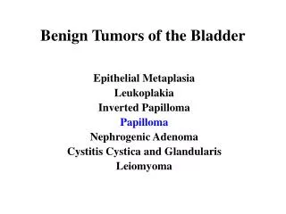
BENIGN TUMORS • Hemangioma (most common) • Liver cell adenoma • Focal nodular hyperplasia • Cystadenoma • Miscellaneous & Rare tumors
HEMANGIOMA • Most common benign tumor of the liver • Female preponderance • Considered as congenital malformation • Usually single and <5cm • >5cm are considered as giant hemangiomas • Microscopically : endothelium lined blood filled spaces separated by thin septae • Usually asymptomatic ,can cause symptoms secondary to compression
Rare complication – rapid expansion,sudden acute thrombosis, sudden spontaneous rupture • Can occur as Kasabach-Merritt syndrome (thrombocytopenia,coagulopathy,hemangioma’s) • Diagnosis – CT/MRI • Management – resection (enucleation with inflow control)
ADENOMA • Rare • Female preponderance • Associated with OCP usage • >10 in number is termed as adenomatosis • Most commonly present as upper abdominal pain • Complications – rupture with fatal intraperitoneal hemorrhage and malignant transformation
Diagnosis – CT(well circumscribed heterogenous mass showing enhancement in arterial phase) , MRI • Management : 1.In near fatal hemorrhage hepatic artery embolization followed by laparotomy and resection 2.In adenomatosis, orthotopic liver transplant is recommended
FOCAL NODULAR HYPERPLASIA • Second most common benign liver tumor • Female preponderance • Usually <5cm involving both lobes of liver • Characterised by central fibrous scar with radiating septa • Considered as developmental vascular malformation • Broadly classified under 2 types classical & non classical(telangiectic,hyperplastic,adenomatous,mixed) • Usually asymptomatic and is a by chance finding on laparotomy
Rare complications – rupture,bleeding,infarction • Diagnosis – CECT/MRI • Management – reassurance but may require resection should symptoms and complications arise
CYSTADENOMA • Rare • Presents as large cystic mass (10-20 cm) • Has globular external surface with multiple protruding cysts & locules • Women > 40years • Can present with abdominal pain,abdomendistention,anorexia and nausea • Can turn into cystadenocarcinoma (rare) • Diagnosis : USG ,CECT/MRI
USG – cystic structure with varying wall thickness,nodularity,septations & fluid filled vacuoles • CECT – enhancement of cyst wall & septae • Management : complete excision must be done as recurrence rate is high (40%)
RARE HEPATIC PATHOLOGIES • Macroregenerative nodules – cirrhotic patients,malignancy potential+ • Nodular regenerative hyperplasia - <2cm,not malignant • Mesenchymalhamartomas – childhood tumor,abdomen mass+, need resection • Fatty tumors of liver – primary lipoma,myelolipoma,angiolipoma,angiomyolipoma
Leiomyoma,myxoma,schwannoma,lymphangioma,teratoma • Benign fibrous tumor of liver • Pseudotumor of liver • Intrahepaticbiliarycystadenoma • Bile duct adenoma • Biliaryhamartoma
THANK YOU -REFERENCES : BAILEY & LOVE(25TH) , SABISTON(18th) -PUBMED CENTRAL (PICTORIAL REFERENCES)