Download
1 / 23
230 likes | 325 Vues
This study aims to develop a knowledge-based system for extracting organs from CT volumes, crucial for patient-specific treatment planning. The system employs dynamic thresholding, shape constraints, and progressive landmarking to tackle challenges in segmenting CT images, such as lack of shape invariants and variations in organ gray tones. Specific properties and constraints tailored to each organ guide the organ identification process step by step. The procedure involves iterative threshold adjustment, shape checks, overlap and collision validations, and selection of the best candidate regions. Morphological operations are applied to extract the final organ of interest. Experimental results are rated based on accuracy grades compared to manual delineation.

E N D
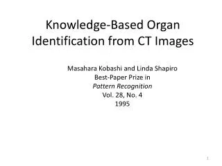
Knowledge-Based Organ Identification from CT Images MasaharaKobashi and Linda Shapiro Best-Paper Prize in Pattern Recognition Vol. 28, No. 4 1995
Motivation • The extraction of structure from CT volumes of cancer • patients is an important first step in the creation of • patient-specific models that can be used by treatment • planning software to deliver maximal dosage to the • tumor and minimal dosage to critical anatomical structures. • Even today, no automatic techniques have been successful • enough to replace the standard manual methods of • outlining the organs. • The goal of this work was to develop a knowledge-based • recognition system that utilizes knowledge of anatomy and • image processing to extract the organs from CT volumes.
3 CT Slices of the Abdomen kidney.jpg g006.jpg e030.jpg Where are the kidneys, liver, spleen, aorta, spine?
Major Features of the System dynamic thresholding controlled by feedback the use of negative shape constraints that 3. progressive landmarking that extracts organs in order of predicted success and uses already-extracted organs to help locate others
Difficulties in Segmenting CT Images Regions produced by gray-tone-based segmentation procedures do not correspond to organs. There are very few shape invariants for organs. The absolute gray tones for each organ vary widely over difference instances. There is no precise, objective ground truth for performance evaluation (in our study).
Observations • Two different organs can have the same or very close • gray tones in CT images • Most human organs have few computable and stable • shape invariants. Shapes of a kidney in different CT slices.
More Observations • Each organ has a fairly stable vertical and horizontal location. • The ordering of organs by their gray tones is fairly stable, • even though their absolute gray tones vary widely. • Each biological substance has a relatively narrow range of • gray tones. • But CT image analysis is simpler than many outside-world • computer vision domains. • And there are relatively small numbers of objects in each • image. • There are some very stable landmarks: spine and aorta.
Specific Organ Properties to Use position in the ordering of gray tones among organs relevant gray-tone range height of gray-tone cliff (related to range of thresholds) location in terms of stable landmarks: aorta and spine adjacency with other organs size in terms of expected area in a slice overlap ratio with other slices positive and negative shape constraints
Idea of the Dynamic Thresholding results of thresholding at the initial (highest) threshold for kidneys at 3 steps, the kidneys become detectible at 11 steps, both kidneys connect with other organs
Steps of the Procedure Set the initial threshold to the high end of the relevant gray tone range for the organ of interest. From the second iteration on, this threshold will be reduced by a constant value (10 was used) in each iteration. If the threshold reaches the low end of the range with no candidates, other methods are invoked.
Steps Threshold the image with the current threshold. Perform connected components to produce a set of regions. AREA CHECK: Check if there is a region of acceptable size in the search area for the organ of interest. If not, go back to step 1. LOCATION CHECK: Check if any candidate regions satisfy the location condition for the organ of interest. If so, record them, else go back to step 1.
Steps SHAPE CHECK: Check if there is among the candidates one that satisfies the positive shape constraints (for the aorta and spine) or the negative shape constraints (for the rest). Negative shape constraints include - abnormal size - abnormal extension - vertical and horizontal lengths - vertical/horizontal ratio
Concept of a Negative Shape Constraint: Shapes that are NOT Kidney
Steps OVERLAP CHECK: check if there is a candidate region that satisfies the overlap condition with an already-segmented adjacent slice. Else go to step 1. The minimum required overlap is 50% of the smaller region. COLLISION CHECK: Check if there is a candidate region that does not collide with other recognized organs. Else go to step 1. 9. CHOOSE BEST: Choose the best candidate region.
Steps SLOPE CHECK: Check the change in area with change in threshold. Look for the flattest part of the curve that has acceptable area. Choose the midpoint as the threshold.
Steps • MORPHOLOGICAL OPERATIONS: • Close with a disk of 3 • Open with a disk of 5 • Extract the regions that satisfies the conditions • Close the extracted region with a disk of 3 • The result is output as the organ of interest.
Some Results: Labeled as Grade A, B, or C Grade A: Comparable to human dosimetry within a 5 pixel mismatch.
Some Results: Labeled as Grade A, B, or C Grade B: Worse than A, but at least 70% correct.
Some Results: Labeled as Grade A, B, or C Grade B Spleen
Some Results: Labeled as Grade A, B, or C Grade C: Less than 70% correct.
Extraction from 3 Slices low-level slice mid-level slice higher-level slice
Possible Course Project • Design and implement a semi-automatic system • that finds and segments organs from CT or other • images and produces 3D meshes from the slices • of each organ: • User: Jim Brinkley in BHI (he’s wanted this forever) We will see this algorithm again used for finding extractible organs in head and neck images in Chia-Chi Teng’s work.