Download
1 / 23
230 likes | 249 Vues
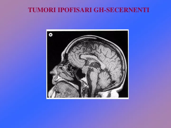
TUMORI IPOFISARI GH-SECERNENTI. ECCESSO DI GH: NON SOLO ACROMEGALIA…. Dalla biologia molecolare…. …alla clinica. Monoclonale. Policlonale. CLONALITA’ DEI TUMORI IPOFISARI.

E N D
Dalla biologia molecolare… …alla clinica.
Monoclonale Policlonale CLONALITA’ DEI TUMORI IPOFISARI - Un tumore può avere una origine MONOCLONALE, che implica il suo sviluppo a partire da una singola cellula geneticamente alterata. - Un tumore può avere una origine POLICLONALE, ossia può originare da un gruppo di cellule stimolate da fattori che ne promuovono la crescita. Tumori ipofisi
Your use of this service is governed by Terms and Conditions. Please review our Privacy Policy for details on how we protect information that you supply.
Voice deepening Headache Snoring , lip, tongue Joint pain
Arachnodactyly (long fingers) Marfan's syndrome and homocystinuria are the two conditions to consider. Of these two, Marfan's syndromeis the most common case you will encounter in the examination. Mention you like to search other physical signs such as tall habitus, arm span longer than height, high-arched palate and kyphosclerosis. Ask to examine the eyes for lens subluxation. The patients may have aphakia due to lens extraction or dislocation. Return to the top Arachnodactyly (long fingers) Marfan's syndrome and homocystinuria are the two conditions to consider. Of these two, Marfan's syndromeis the most common case you will encounter in the examination. Mention you like to search other physical signs such as tall habitus, arm span longer than height, high-arched palate and kyphosclerosis. Ask to examine the eyes for lens subluxation. The patients may have aphakia due to lens extraction or dislocation. Return to the top Polydactyly (extract digit) There are many conditions associated with polydactyly. However, onlyLaurence-Moon's and Biedl-Bardet's Syndromes are likely to appear inthe examination. The extract digits may not be immediately apparent unless you count them. In some patients, the digits may have been amputated in childhood leaving behind only scar(s). Mention that you would like to examine the fundi for any evidence of pigmentary retinopathy. The examination may be difficult due to a lack of cooperation as these patients may have associated mental handicap. Return to the top Polydactyly (extract digit) There are many conditions associated with polydactyly. However, onlyLaurence-Moon's and Biedl-Bardet's Syndromes are likely to appear inthe examination. The extract digits may not be immediately apparent unless you count them. In some patients, the digits may have been amputated in childhood leaving behind only scar(s). Mention that you would like to examine the fundi for any evidence of pigmentary retinopathy. The examination may be difficult due to a lack of cooperation as these patients may have associated mental handicap. Return to the top Large hands The hands are large and the fingers are broad. The skin on the dorsum of the hands is thickened (demonstrated by gently pinching the skin).The most likely case is acromegaly. Mention that you like to examine the visual field of the patient for any evidence of bitemporal hemianopia. Also look for any optic atrophy which is classically described as bow-tie atrophy. Return to the top Large hands The hands are large and the fingers are broad. The skin on the dorsum of the hands is thickened (demonstrated by gently pinching the skin).The most likely case is acromegaly. Mention that you like to examine the visual field of the patient for any evidence of bitemporal hemianopia. Also look for any optic atrophy which is classically described as bow-tie atrophy. Return to the top • Rheumatoid arthritis • The hands show symmetrical arthropathy, consisting of:swelling of the phalangeal joints except the distal phalangeal joints (the swelling is caused by synovial swelling) • Z-deformity of the thumb • Boutonniere deformity • swan neck deformity • ulnar deviation of metacarpophalangeal joint • volar subluxation of the palm • The palmar sides show palmar erythema and wasting of the muscles. • The muscle power may be weak due to compression of thenerve or rupture of the tendon. The function of the hands such as writing and unbuttoning are usually good despitethe gross deformity. • Check for the following signs: • rheumatoid nodules at the elbows • scars at the wrist from operation for carpal tunnel syndrome • associated ocular signs: such as dry eyes and scleromalacia perforans • Return to the top • Rheumatoid arthritis • The hands show symmetrical arthropathy, consisting of:swelling of the phalangeal joints except the distal phalangeal joints (the swelling is caused by synovial swelling) • Z-deformity of the thumb • Boutonniere deformity • swan neck deformity • ulnar deviation of metacarpophalangeal joint • volar subluxation of the palm • The palmar sides show palmar erythema and wasting of the muscles. • The muscle power may be weak due to compression of thenerve or rupture of the tendon. The function of the hands such as writing and unbuttoning are usually good despitethe gross deformity. • Check for the following signs: • rheumatoid nodules at the elbows • scars at the wrist from operation for carpal tunnel syndrome • associated ocular signs: such as dry eyes and scleromalacia perforans • Return to the top • Psoriatic arthropathy • The nails show pitting and onycholysis (lifting of the nail fromthe nail bed). There are swellings of the joints involving the distal interphalangeal joint (contrast this with rheumatoid arthritis in which the distal phalangeal joints are not involved). Sometimesthe joint involvement may resemble that of rheumatoid arthritis. Rarely do you get arthritis mutilans (telescoping of phalanges). The function of the hands is usually good unless the joints are severely inflamed. • Check for the following signs: • look for psoriatic plaques (which may not be apparent on the hands as in the picture here, they have well-defined edges with whitish scales) at the elbow, knees and behind the ears • mention you would like to examine the eyes on the slit-lamp for anterior uveitis or signs of past inflammation such as pigments on the lens or posterior synechiae. • Return to the top • Psoriatic arthropathy • The nails show pitting and onycholysis (lifting of the nail fromthe nail bed). There are swellings of the joints involving the distal interphalangeal joint (contrast this with rheumatoid arthritis in which the distal phalangeal joints are not involved). Sometimesthe joint involvement may resemble that of rheumatoid arthritis. Rarely do you get arthritis mutilans (telescoping of phalanges). The function of the hands is usually good unless the joints are severely inflamed. • Check for the following signs: • look for psoriatic plaques (which may not be apparent on the hands as in the picture here, they have well-defined edges with whitish scales) at the elbow, knees and behind the ears • mention you would like to examine the eyes on the slit-lamp for anterior uveitis or signs of past inflammation such as pigments on the lens or posterior synechiae. • Return to the top
ACROMEGALYCushing: The pituitary body & its disorders Before disease Disease onset 12 yrs of disease 17 yrs of disease Square hand Phalanges
Diagnosi Inizio della malattia
MORTALITA’ NELL’ACROMEGALIA La mortalità nel paziente acromegalico è circa il doppio rispetto a quella attesa nella popolazione sana. La mortalità è legata soprattutto a: Malattie cardio-vascolari Malattie respiratorie Malattie cerebro-vascolari Malattie neoplastiche
TERAPIA DELL’ACROMEGALIA La terapia degli adenomi ipofisari GH-secernenti può essere chirurgica, radioterapica o medica. Essa è finalizzata a: a) rimuovere la neoplasia o ridurne il volume; b) sopprimere l'ipersecrezione ormonale; c) risolvere o almeno limitare insorgenza e progressione delle complicanze; d) correggere eventuali deficit ipofisari associati mediante adeguato trattamento sostitutivo.
Guarigione = • GH < 2.5 ng/ml • Normalizzazione IGF-1 • GH (nadir) < 1 (0.3) ng/ml dopo OGTT Melmed et al., J Clin Endocrinol Metab 5 1509-1517, 2009 TERAPIA CHIRURGICA Rappresenta tuttora il trattamento di prima scelta. Approccio per via trans-sfenoidale. Guarigione > 80% dei casi se microadenoma (< 1 cm diametro), < 50% se macroadenoma (> 1 cm diametro)
TERAPIA RADIANTE - La radioterapia è stata per lungo tempo considerata l’opzione terapeutica di seconda scelta e generalmente proposta nelle situazioni in cui le condizioni del paziente non consentano l’intervento o se il tumore è profondamente infiltrato a livello della regione ipotalamica. - La radioterapia può essere effettuata con metodica convenzionale, frazionata a campi contrapposti o stereotassica. - Il limite principale di quest’opzione terapeutica è rappresentato dall’efficacia assai tardiva (anche 10 anni o più) e dalle possibili complicanze (es. frequente ipopituitarismo). - Oggi è spesso opzione di terza scelta.
TERAPIA FARMACOLOGICA Utilizzata come trattamento di “seconda linea” (dopo chirurgia o in attesa di risultato della radioterapia). • Tre classi di farmaci disponibili • Analoghi della somatostatina (octreotide, lanreotide) (successo 60/70% pz.) • Dopamino-agonisti (successo < 10% pz). • Antagonisti recettore GH (pegvisomant). Quest’ultimo indicato per pazienti resistenti agli altri trattamenti (circa 30%).
La terapia medica può essere trattamento di scelta in casi in cui le dimensioni e l’estensione sia tale da non rendere possibile un successo chirurgico o in presenza di controindicazioni alla chirurgia legate ad età e/o comorbidità del paziente.