Download

1 / 15
160 likes | 218 Vues
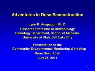
Explore the criticality accident in Tokaimura, Japan, and the cytogenetic dose estimation methods used. Learn about the accident's chronology, blood cell counts, and chromosome preparations post-accident.

E N D
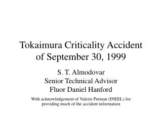
Cytogenetic Dose Estimation in the Criticality Accident in Tokaimura Lecture Module 13
Criticality Accident in Tokai-mura, Japan • On 30 September 1999, at 10:35, a criticality accident occurred at the uranium conversion facility in Tokai-mura, a village 130 km northeast from Tokyo, Ibaraki Prefecture, Japan • The criticality event occurred when a worker was pouring a solution of enriched uranium (235U) into a precipitation tank directly • He bypassed a dissolution tank and buffer column supposed to be used in order to avoid criticality • The amount of uranium poured was several times more than the specified mass limit
Where did accident occur? Tokai-mura The JCO is located at Tokai-mura, Ibaraki Prefecture, which is about 130 km northeast of Tokyo.
Chronology of accident loss of consciousness JCO NIRS
Blood Cells Counts 2-3 h after accident From J Radiat Res 2001 42 Suppl S157-166
Chromosome Preparations Scoring Dic+R Centrifugal sedimantation RPMI-1640 20% FCS 2% PHA 37ºC 48h incubate Add 75mM KCL 37ºC 20min 0.3μg Colcemid Fixed with 1:3 acetic alcohol (three times) Mononucleated cells -20ºC 3h 37ºC 48h incubate 8ml peripheral blood Air-dry slides 500nM Okdaic acid (last 1h) PCC-ring analysis
Prematurely condensed chromosomes having PCC-rings (white arrow) in a lymphocyte of patient A First Performed PCC-ring Analysis
Metaphase Chromosomes in Patient B Metaphase chromosomes having dicentric chromosomes (black arrows), a tricentric chromosome (short arrow) and a ring chromosome with centromere (white arrow) in a lymphocyte of patient B
Metaphase Chromosomes in Patient A Metaphase chromosomes having severe chromosome aberrations in a lymphocyte of patient A
Frequency of Chromosome Aberration in Lymphocytes Dic: dicentric chromosome R: ring chromosome with/without centromere Rc: ring chromosome with centromere
Dose-response Curves Dose-response curve (Y=2.31 x 10⁻²D + 6.33 x 10⁻²D2) of Dic+Rc for 60Co γ-rays, dose-response curves of Dic+R, Dic+Rc, and Dic for 1.9 MeV x-rays (after Norman and Sasaki, 1966), and the estimated dose of patients A (□) and B (∆).
Comparison of Doses Estimated by Various Indicators * Equivalent dose to X or γ-rays ** Ishigure et al, when neutoron’s RBE is 1.5-2.0.
Conclusion • It was difficult to collect sufficient number of lymphocytes due to severely high dose exposure • High-yield chromosome preparation method was used to collect lymphocytes • Dose estimation was made by two method: • PCC-ring analysis • Analysis of dicentric and ring chromosome