Toxic Epidermal Necrolysis
230 likes | 689 Vues
Toxic Epidermal Necrolysis. Kristine Scruggs, MD AM Report July 28, 2009. Definition. SJS/TEN: Lesions: Small blisters on dusky purpuric macules or atypical targets Mucosal involvement common Prodrome of fever and malaise common Stevens-Johnson Syndrome: Rare areas of confluence.

Toxic Epidermal Necrolysis
E N D
Presentation Transcript
Toxic Epidermal Necrolysis Kristine Scruggs, MD AM Report July 28, 2009
Definition • SJS/TEN: • Lesions: Small blisters on dusky purpuric macules or atypical targets • Mucosal involvement common • Prodrome of fever and malaise common • Stevens-Johnson Syndrome: • Rare areas of confluence. • Detachment </= 10% BSA • Toxic Epidermal Necrolysis: • Confluent erythema is common. • Outer layer of epidermis separates easily from basal layer with lateral pressure. • Large sheet of necrotic epidermis often present. • >30% BSA involved.
Presentation • Fever (often >39) and flu-like illness 1-3 days before mucocutaneous lesions appear • Confluent erythema • Facial edema or central facial involvement • Lesions are painful • Palpable purpura • Skin necrosis, blisters and/or epidermal detachment • Mucous membrane erosions/crusting, sore throat • Visual Impairment (secondary to ocular involvement) • Rash 1-3 weeks after exposure, or days after 2nd exposure
2-7/million people/year SJS: age 25-47, TEN: age 46-63 Women: >60% Poor prognosis: Intestinal/Pulmonary involvement Greater extent of detachment Older age Mortality: SJS: 5% TEN: 30% Risk Factors: HIV infection Genetic factors Certain HLA types “Slow acetylators” Polymorphisms in IL4 receptor gene Concomitant viral infections Underlying immunologic diseases Physical factors UV light, radiation therapy Malignancy Higher doses of known offenders Epidemiology
Pathogenesis • Secondary to cytotoxicity and delayed hypersensitivity reaction to the offending agent. • Antigen is either the implicated drug or a metabolite. • Histopathology: • Granulysin (cytolytic protein produced by cytotoxic T cells and NK cells) • Expression of HLA-DR and intracellular adhesion molecule (ICAM)-1 by • Keratinocytes • CD4 cells (in dermis) • CD8 T cells (in epidermis) • Apoptosis of keratinocytes facilitated by • TNF-alpha, perforin and granzyme secretion • fas-ligand expression (cell death receptor) Subepidermal split with cell-poor bullous. Epidermis shows full thickness necrosis.
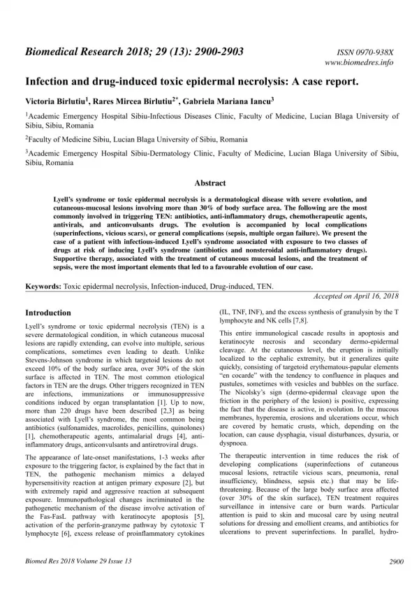
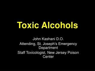
Etiologies • Medications (Odds Ratio for exposure in hospitalized pts): • Sulfonamide antibiotics (172) • Allopurinol (52) • Amine antiepileptics • Phenytoin (53) • Carbamazepine (90) • Lamotrigine • NSAIDs (72) • Infections (e.g. Mycoplasma pneumonia) • Other: Vaccinations, Systemic diseases, Chemical exposure, Herbal medicines, Foods
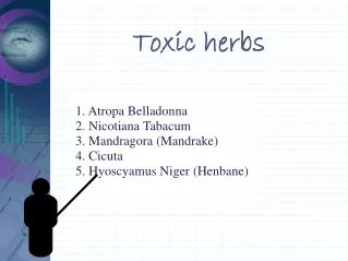
Differential Diagnosis for Vesicular or Bullous Rash Bullous Pemphigoid Often affects the elderly Dermatitis Herpetiformis Associated with gluten intolerance Pemphigus Affects middle-aged or elderly Cicatricial Pemphigoid Mucosal involvement, sometimes cutaneous
Differential Diagnosis, cont. Linear IgA Disease Itchy, ring-shaped, no internal disease Herpes Simplex Virus Varicella/Zoster Virus Hand-Foot-Mouth Disease (Enteroviruses) Contact Dermatitis
Differential Diagnosis, cont. • Erythema Multiforme • Staphylococcal Scalded Skin Syndrome • Bullous Impetigo • Toxic Shock Syndrome • Paraneoplastic Pemphigus • Cutaneous emboli • Diabetic Bullae • Porphyria Cutanea Tarda • Porphyria Variegata • Pseudoporphyria • Bullous dermatosis of Hemodialysis • Coma Bulloae • Epidermolysis Bullosa Acquisita
Treatment • Early diagnosis - biopsy • Immediate discontinuation of offending agent • Supportive care – pay close attention to ocular complications • IV hydration (e.g. Parkland formula) • Antihistamines • Analgesics • Local v. systemic corticosteroids • Think about nursing requirements! • Possible treatment in burn unit, wound care • IVIg?
Resources: • Cooper, et al. The Washington Manual of Medical Therapeutics, 32nd Edition. 2007. • High, et al. Stevens-Johnson syndrome and toxic epidermal necrolysis: Management, prognosis, and long-term sequelae. Up To Date. 2009. • Kasper, et al. Harrison’s Principles of Internal Medicine, 16th Edition. 2005. • Nirken, et al. Stevens-Johnson syndrome and toxic epidermal necrolysis: Clinical manifestations, pathogenesis, and diagnosis. Up To Date. 2009.