Download
1 / 17
400 likes | 3.96k Vues
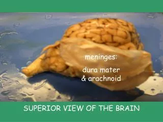
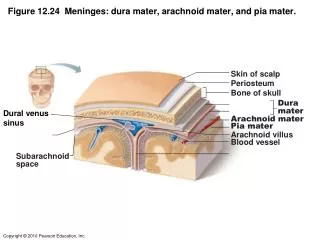
Figure 12.24 Meninges: dura mater, arachnoid mater, and pia mater. Skin of scalp. Periosteum. Bone of skull. Dura mater. Dural venus sinus. Arachnoid mater. Pia mater. Arachnoid villus. Blood vessel. Subarachnoid space. Figure 11.15 The brain. Midbrain. Figure 11.15 The brain.

E N D
Figure 12.24 Meninges: dura mater, arachnoid mater, and pia mater. Skin of scalp Periosteum Bone of skull Dura mater Dural venus sinus Arachnoid mater Pia mater Arachnoidvillus Blood vessel Subarachnoid space
Figure 11.15 The brain. Midbrain
Figure 11.15 The brain. FOREBRAIN HINDBRAIN
Figure 12.12 Midsagittal section of the brain illustrating the diencephalon (purple) and brain stem (green). Lateral ventricle Corpus callosum Fornix Choroid plexus Thalamus (encloses third ventricle) Pineal gland Mid- brain Hypothalamus Optic chiasma Arbor vitae (of cerebellum) Pituitary gland Fourth ventricle Mammillary body Choroid plexus Pons Cerebellum Medulla oblongata Spinal cord
Hormone-producing organs of the brain • Hypothalamus • Pituitary gland • Pineal grand
Figure 12.14 Inferior view of the brain Frontal lobe Olfactory bulb Optic chiasma Mammillary body Midbrain Pons Temporal lobe Medulla oblongata Cerebellum Spinal cord
Functions of cerebellum • Regulates balance and posture, by processing information from visual and equilibrium pathways • Works with cerebrum to provide smooth, coordinated skeletal muscle movements
Figure 12.17a Cerebellum. Arbor vitae Pons Fourth ventricle Medulla oblongata Choroid plexus (a)
Pattern of gray and white matter in the embryonic spinal cord, brain stem and cerebellum. Cortex of gray matter Central cavity Inner gray matter Outer white matter Gray matter Region of cerebellum Central cavity Inner gray matter Outer white matter Gray matter Brain stem Central cavity Outer white matter Inner gray matter Spinal cord
Figure 11.13 The ventricles of the brain and circulation of cerebrospinal fluid.
Figure 11.13 The ventricles of the brain and circulation of cerebrospinal fluid.
Figure 12.26a Formation, location, and circulation of CSF. Dural venous sinus 3 Choroid plexus Arachnoid villus Subarachnoid space Meningeal dura mater 1 Right lateral ventricle (deep to cut) Choroid plexus 2 Third ventricle 1. CSF is produced by the choroid plexus of each ventricle. Cerebral aqueduct Fourth ventricle 2. CSF flows through the ventricles and into the subarachnoid space. Some CSF flows through the central canal of the spinal cord. 2 Central canal of spinal cord 3. CSF is absorbed into the dural venous sinuses via the arachnoidvilli.
Figure 12.26b. CSF formation by choroid plexuses Ependymal cells Capillary Section of choroid plexus CSF forms as a filtrate containing glucose, oxygen, vitamins, and ions (Na+, Cl–, Mg2+, etc.) Cavity of ventricle
Figure 11.16 The cerebrum. Longitudinal fissure
Figure 12.6a Some lobes and fissures of the cerebral hemispheres Precentralgyrus Central sulcus Postcentralgyrus Frontal lobe Parietal lobe Parieto-occipital sulcus Lateral sulcus Occipital lobe Temporal lobe Fissure (a deep sulcus) Gyrus Cortex (gray matter) Sulcus White matter
Figure 11.17 Primary somatosensory and motor areas of the cerebral cortex.
Figure 11.21 Changes in brain metabolic activity in Alzheimer’s disease.