The Reproductive System Functions and Processes
870 likes | 917 Vues
Explore the male and female reproductive systems, cell division, gametogenesis, hormones, and key anatomical structures. Learn about spermatogenesis, spermiogenesis, oocyte maturation, and more.

The Reproductive System Functions and Processes
E N D
Presentation Transcript
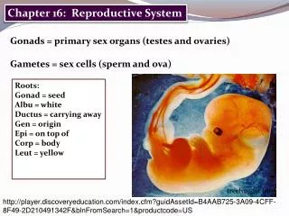
Chapter 28: The Reproductive System BIO 211 Lecture Instructor: Dr. Gollwitzer
Today in class we will: • Compare the male and female reproductive systems • Discuss cell division • Compare and contrast mitosis and meiosis • Discuss gametogenesis (spermatogenesis) in the male • Spermiogenesis • Spermiation • Capacitation • Anatomy of a spermatozoon • Structures involved in spermiogenesis and their roles • Describe the composition of seminal fluid • Identify the glands whose secretions contribute to the production of seminal fluid • Discuss male reproductive endocrinology • Endocrine structures and hormones that regulate male reproductive function
Reproductive System • Only organ system not essential to life • Ensures continued existence of human species • Produces, stores, nourishes, and transports male and female reproductive cells (gametes) • Produces reproductive hormones
Male and Female Reproductive Systems • Functionally very different • Female produces 1 gamete/month • Retains and nurtures zygote • Male produces large quantities of gametes • 500M/day! • Begins at puberty and continues past age 70
Male • Testes (male gonads) • Produce male gametes (spermatozoa, sperm) • Produce hormones • Male sex hormones (androgens, primarily testosterone) • Inhibin • Emission • Movement of mature spermatozoa move through male duct system, are mixed with secretions of accessory glands • Semen • Sperm mixed with accessory gland secretions
Female • Ovaries (female gonads) • Release 1 immature gamete (oocyte) each month • Produce hormones • Female sex hormones (estrogens, progestins) • Inhibin • Uterine tube carries oocyte to uterus • If sperm reaches oocyte in uterine tube: • Fertilization is initiated • Oocyte matures into ovum
Reproduction • During sexual intercourse, ejaculation introduces semen into vagina • Spermatozoa ascend female reproductive tract • Seek out oocyte (generates heat, attracts sperm like heat-seeking missile) • If fertilization occurs in uterine tube: • sperm + ovum zygote • Zygote travels to uterus • Uterus encloses/supports developing embryo • Embryo grows into fetus and prepares for birth
Gametogenesis • Involves mitosis and meiosis • Mitosis • Process of somatic cell division • Produces 2 diploid daughter cells • Have same number of (paired) chromosomes as parent cell, i.e., 46 (23 x 2) • Meiosis = reduction division • Special cell division involved in gamete production • Produces 2 haploid daughter cells • Have one-half (unpaired) the number of chromosomes in the parent cell, i.e., 23
Chromosomes in Mitosis and Meiosis Figure 28–6
Gametogenesis • Meiosis • Involves two cycles of cell division • Chromosomes (each with two chromatids) pair up = tetrad • During first division, tetrads split • During second division, chromatids split • Produces gametes with one-half the number of chromosomes, i.e., 23 • Fusion of male gamete (sperm) and female gamete (oocyte) produces cell with correct number of chromosomes (diploid), i.e., 46 (23 from each parent)
Spermatogenesis • Occurs in seminiferous tubules (ST) in testes • 3 integrated processes • Mitosis • Meiosis • Spermiogenesis
Spermatogenesis • Mitosis • Spermatogonium (stem cell) spermatogonium + primary spermatocyte • Primary spermatocyte pushed toward lumen of ST • On-going throughout lifetime • Meiosis • Primary spermatocyte first division secondary spermatocytes second division spermatids = undifferentiated male gametes • Each primary spermatocyte 4 spermatids
Spermatogenesis • Spermiogenesis • Last stage of spermatogenesis • Begins with spermatids • Small, relatively unspecialized cells • Physical maturation of spermatids • Involves major structural changes • Differentiate into mature spermatozoa • Highly specialized cells
Spermiation • When spermatozoa: • Detach from Sertoli cells • Enter lumen of ST • From spermatogonium to spermiation: • 9 weeks
Seminiferous Tubules Figure 28–5a
Seminiferous Tubules Figure 28–5d
Spermiogenesis and Spermatozoon Structure Figure 28–8
Anatomy of a Spermatozoon • Head – nucleus with chromosomes (DNA) • Acrosomal cap – contains enzymes to dissolve oocyte wall • Middle piece – contains mitochondria for energy to move tail • Tail – flagellum (only 1 in human body); provides motility • Loses all other organelles to make light weight • No energy reserves – must use nutrients from surrounding fluid (primarily fructose)
Interstitial (Leydig) Cells • Large cells in interstitial spaces between ST • Stimulated by LH androgens (testosterone, T) • Testosterone • Stimulates spermatogenesis and spermatozoa maturation • Affects CNS, including libido (sexual drive) • Stimulates metabolism, especially protein synthesis, muscle growth • Establishes/maintains secondary sex characteristics, e.g., facial hair • Maintains male accessory glands and organs
Seminiferous Tubules Figure 28–5b,c
Sustentacular (Sertoli) Cells • “Nurse cells” • Extend between other cells from ST capsule to lumen • Surround developing spermatocytes and spermatids in ST • 6 major functions • Maintain blood-testis barrier • Support mitosis and meiosis • Support spermiogenesis • Produce inhibin • Produce androgen-binding protein (ABP) • Secrete Mullerian-inhibiting factor (MIF)
Sustentacular (Sertoli) Cells • Maintain blood-testis barrier • Cells joined by tight junctions • Isolates STs • Support mitosis and meiosis • Cells stimulated by FSH (and presence of T) • Promote spermatogenesis • Support spermiogenesis • Provide nutrients for development • Phagocytize cytoplasm shed by spermatids
Sustentacular (Sertoli) Cells • Produce inhibin • Stimulated by factors released by developing spermatozoa • Provides feedback control of spermatogenesis • Inhibits (decreases) production of FSH by AP • Produce androgen-bind protein (ABP) • Stimulated by FSH • Binds T in ST fluid, elevates levels • Produce Mullerian-inhibiting factor (MIF) • Causes regression of fetal ducts that form uterine tubes and uterus
Epididymis • Spermatozoa in ST functionally immature • Incapable of fertilization or locomotion • Become functionally mature in epididymis (but, not motile) • Fluid currents (from cilia lining efferent ductules) transport immobile gametes into epididymis • Functions • Monitors and adjusts composition of ST fluid • Recycles damaged spermatozoa • Stores/protects spermatozoa and facilitates functional maturation • Transit time = two weeks
NOTE: • To become motile, spermatozoa must undergo capacitation • Become motile when mixed with seminal vesicle fluid • Capable of successful fertilization when exposed to female reproductive tract
Ductus (Vas) Deferens • Transport spermatozoa from epididymis to urethra • Store spermatozoa (several months) • In state of suspended animation • Low metabolic rates
Seminal Fluid • A mixture of secretions from several glands including: • Seminal vesicles (60%) • Prostate gland (20-30%) • Bulbourethral glands (10-20%)
Seminal Vesicles • Secretions contain: • High concentrations of fructose (easily metabolized by spermatozoa) • Prostaglandins – stimulate smooth muscle contractions in male and female reproductive tracts • Fibrinogen – forms temporary clot in vagina after ejaculation (seminal plug) • Secretions make functional spermatozoa motile (flagella begins beating) • Secretions discharged into ejaculatory duct at emission (due to contractions in ductus deferens, SVs, and prostate gland)
Prostate Gland • Produces prostatic fluid • Contains seminalplasmin = antibiotic that may help prevent urinary tract infections • Ejected into prostatic urethra
Bulbourethral (Cowper’s) Glands • Mucous glands • Secretions • Help neutralize urinary acids remaining in urethra • Lubricate glans (tip of) penis
Semen • Ejaculate = 2-5 mL of semen • Contains • Spermatozoa • Sperm count = 20 – 100 million/mL semen (ideally > 60 million/ejaculate) • Seminal fluid = mixture of glandular secretions from: • SV (60%) • Prostate (30%) • Bulbourethral glands (5%) • Sustentacular cells and epididymis (5%) • Enzymes • Protease – helps dissolve vaginal mucous secretions • Seminalplasmin (from prostate) • Prostatic enzyme - converts fibrinogen to fibrin after ejaculation • Fibrinolysin – liquefies clotted semen
Male Reproductive Endocrinology • Hypothalamus GnRH ant pit • Ant pit • LH (ICSH) interstitial (Leydig) Cells testosterone (T) • FSH • Testosterone + FSH sustentacular cells • Synthesis of ABP • Stimulation of spermatogenesis and spermiogenesis • Factors released by developing spermatozoa sustentacular cells inhibin inhibits (decreases) FSH production by AP
Physiological Effects of Testosterone • Stimulates spermatogenesis (with FSH) • Maintains male accessory glands and organs • Establishes/maintains secondary sex characteristics • Stimulates anabolic metabolism, especially bone and muscle growth, RBC formation • On CNS, including libido (sexual drive)
Male Reproductive Endocrinology • In males, GnRH pulse frequency relatively steady narrow range of plasma FSH, LH, T • T secretion accelerates at puberty • Sexual maturation • Appearance of secondary sex characteristics • Negative feedback controls T production • Inc T inhibits release of GnRH dec LH dec T
Today in class we will discuss: • The female reproductive system • Gametogenesis (oogenesis) in the female • Compare oogenesis to spermatogenesis • The events and structural changes associated with the ovarian cycle • The structure and histology of the uterus • The events and structural changes associated with the uterine (menstrual) cycle • Discuss female reproductive endocrinology • Endocrine structures and hormones that regulate female reproductive function • Aging and the reproductive system of males and females
Female Reproductive System • Produces gametes and reproductive hormones • Protects and supports developing embryo • Nourishes newborn infant
Oogenesis • = Ovum production • Begins before birth, accelerates at puberty, ends at menopause • Occurs monthly between puberty and menopause
Oogenesis vs. Spermatogenesis • Before birth • Mitotic divisions complete • Oogonia (stem cells) primary oocytes (vs. ongoing throughout lifetime in males) • Primary oocytes begin meiosis I • Cytoplasm of oocyte unevenly distributed during 2 meiotic divisions; produces: • One functional ovum (with most of original cytoplasm) • 2-3 polar bodies = nonfunctional cells that later disintegrate (vs. primary spermatocyte 4 spermatozoa) • Ovary releases secondary oocyte instead of mature ovum • Meiosis not completed unless/until fertilization
Primary Oocytes • Are daughter cells of oogonia (stem cells) • Located in ovarian cortex in clusters (egg nests) • Surrounded by follicle cells = primordial follicle • 2 M primary oocytes at birth • Meiosis I (primary oocyte secondary oocyte) • Begins during fetal development • Stops early in meiosis I (suspended development) • Doesn’t continue until after puberty (numbers reduced to 400,000 due to atresia)
Ovarian Cycle • Begins after puberty when groups of primordial follicles develop into primary follicles each month • Process begins due to increased FSH at puberty • Involves 2 phases • Follicular (preovulatory) phase • Luteal (postovulatory) phase
Ovarian Cycle Figure 28-16
Ovarian Cycle: Follicular Phase • Formation of primary follicles • Every month AP FSH some primordial follicles (with primary oocyte) primary follicles (with primary oocyte) • Zona pellucida = glycoprotein layer around primary oocyte • Granulosa cells = rounded, larger follicle cells outside zona pellucida • Thecal cells = layer of follicle cells adjacent to ovarian stroma • Granulosa + thecal cells work together estrogen (continues in all follicles)
Ovarian Cycle: Follicular Phase • Formation of secondary follicles • Each month, only a few (1-3) primary follicles become secondary follicles • Primary oocyte increases in size • Follicle wall thickens • Granulosa cells secrete follicular fluid • Follicle enlarges rapidly as follicular fluid accumulates