Download
1 / 36
360 likes | 382 Vues
Learn about the risk factors, pathophysiology, clinical presentation, and diagnostic tests for infective endocarditis. Explore treatment options and preventive measures.

E N D
Infectious Disease I:Infective Endocarditis Courses in Therapeutics and Disease State Management
Learning Objectives (Slide 1 of 3) • List patient populations at increased risk for developing infective endocarditis (IE) • Delineate bacteria that commonly cause IE as well as situations where certain bacteria are more likely • Describe the sequential steps necessary to develop hematogenous spread of IE • Identify the clinical manifestations of the disease, including physical findings, laboratory abnormalities, blood cultures, and other diagnostic test (e.g., echocardiography) • Argue the importance of correctly obtained blood cultures and state situations that may lead to “culture-negative” IE
Learning Objectives (Slide 2 of 3) • Justify the rationale for high-dose parenteral, bactericidal, extended-duration antibiotics for IE treatment • Summarize the role of nonpharmacologic approaches (i.e., surgery) in the treatment of IE and identify situations where this approach is preferred • Design drug regimens for the following types of infective endocarditis: streptococci, staphylococci, enterococci, the HACEK microorganisms, and “culture-negative” IE • Describe why β-lactam antibiotics are preferred for the treatment of IE and classify situations where vancomycin is appropriate • Evaluate the role of penicillin skin tests in patients with a documented penicillin allergy
Learning Objectives (Slide 3 of 3) • Outline specific monitoring parameters during IE treatment, including signs and symptoms, blood cultures, microbiologic tests, serum drug concentrations, and tests that evaluate organ function • Identify patients who should receive antimicrobials for IE prophylaxis as well as bacteremia-causing procedures that can lead to IE in predisposed individuals • In high-risk groups receiving bacteremia-causing procedures, devise a prophylactic antimicrobial regimen and list alternative regimens in those with an immediate-type penicillin allergy
Required Reading • Veverka A, Crouch MA, Odle BL. Chapter 89. Infective Endocarditis. In: DiPiro JT, Talbert RL, Yee GC, Matzke GR, Wells BG, Posey L. eds. Pharmacotherapy: A Pathophysiologic Approach, 9e. New York, NY: McGraw-Hill; 2014. • Baddour LM, Wilson WR, Bayer AS, et al. Infective endocarditis in adults: diagnosis, antimicrobial therapy, and management of complications: a scientific statement for healthcare professionals from the American Heart Association. Circulation. 2015; 132:1435–86. • Gerber MA, Baltimore RS, Eaton CB, et. Al. Prevention of rheumatic fever and diagnosis and treatment of acute Streptococcal pharyngitis: a scientific statement from the American Heart Association Rheumatic Fever, Endocarditis, and Kawasaki Disease Committee of the Council on Cardiovascular Disease in the Young, the Interdisciplinary Council on Functional Genomics and Translational Biology, and the Interdisciplinary Council on Quality of Care and Outcomes Research. Circulation. 2009;119:1541–15
Overview • Serious infection involving the lining and valves of the heart • Acute Disease • High fevers • Elevated WBC counts • Systemic toxicity • Sub-acute Disease • Slower and more subtle presentation • Low grade fevers • Night sweats • Fatigue
Risk Factors • Presence of a prosthetic valve (highest risk) • Previous endocarditis (highest risk) • Congenital heart disease (CHD) • Chronic IV access • Diabetes mellitus • Healthcare-related exposure • Acquired valvular dysfunction • Cardiac implantable device • Chronic heart failure • Mitral valve prolapse with regurgitation • IV drug abuse
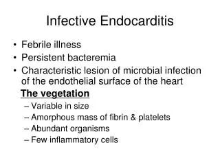
Pathophysiology • Hematogenous spread is the most common pathway • Endothelial surface of the heart must be damages • Platelet and fibrin depositions occur on the damaged epithelial surface • Bacteremia gives organisms access to and results in colonization of the endocardial surface • After colonization of the endothelial surface, a “vegetation” of fibrin, platelets, and bacteria forms • Implantation of prosthetic values or other cardiac hardware that has been contaminated with pathogens is another pathway for endocarditis
Clinical Presentation (Slide 1 of 3) Symptoms Signs Fever New or changing heart murmur Embolic Phenomena Skin manifestations Clubbing of extremities • Fever • Chills • Night Sweats • Weakness • Dyspnea • Weight Loss • Myalgia or arthralgia
Clinical Presentation (Slide 2 of 3) Laboratory Tests Diagnostic Tests Electrocardiogram Chest radiograph Echocardiogram Transthoracic (TTE) Transesophogeal (TEE) • WBC count normal or elevated • Anemia • Elevated C-reactive protein (CRP) • Elevated erythrocyte sedimentation rate (ESR) • Altered urinary analysis • Blood Cultures
Clinical Presentation (Slide 3 of 3) • The signs and symptoms of infective endocarditis are not specific, and the diagnosis is often unclear • The Duke diagnostic criteria integrate clinical, laboratory, and echocardiographic findings to identify the likelihood a patient has endocarditis • Patients are grouped into one of three categories • Definite infective endocarditis • Possible infective endocarditis • Infective endocarditis rejected
Modified Duke Criteria:Major Criteria (Slide 1 of 2) • Blood culture positive for IE • Typical microorganisms consistent with IE from 2 separate blood cultures: Viridans streptococci, Streptococcus bovis, HACEK group, Staphylococcus aureus; or community-acquired enterococci in the absence of a primary focus; or • Microorganisms consistent with IE from persistently positive blood cultures defined as follows: At least 2 positive cultures of blood samples drawn 12 h apart; or all of 3 or a majority of 4 separate cultures of blood (with first and last sample drawn at least 1 h apart) • Single positive blood culture for Coxiella burnetii or anti–phase 1 IgG antibody titer >1:800
Modified Duke Criteria:Major Criteria (Slide 2 of 2) Evidence of endocardial involvement Echocardiogram positive for IE (TEE recommended for patients with prosthetic valves, rated at least “possible IE” by clinical criteria, or complicated IE paravalvular abscess; TTE as first test in other patients) defined as follows: oscillating intracardiac mass on valve or supporting structures, in the path of regurgitant jets, or on implanted material in the absence of an alternative anatomic explanation; or abscess; or new partial dehiscence of prosthetic valve; new valvular regurgitation (worsening or changing or preexisting murmur not sufficient)
Modified Duke Criteria:Minor Criteria Predisposition, predisposing heart condition, or IVDA Fever, temperature >38°C Vascular phenomena, major arterial emboli, septic pulmonary infarcts, mycotic aneurysm, intracranial hemorrhage, conjunctival hemorrhages, and Janeway’s lesions Immunologic phenomena: glomerulonephritis, Osler’s nodes, Roth’s spots, and rheumatoid factor Microbiological evidence: positive blood culture but does not meet a major criterion as noted above* or serological evidence of active infection with organism consistent with IE
Modified Duke Criteria:Diagnostic Scoring (Slide 1 of 2) • Definite Infective Endocarditis • Pathological criteria • Microorganisms demonstrated by culture or histological examination of a vegetation, a vegetation that has embolized, or an intracardiac abscess specimen • Pathological lesions; vegetation or intracardiac abscess confirmed by histological examination showing active endocarditis • Clinical criteria • 2 major criteria • 1 major criterion and 3 minor criteria • 5 minor criteria
Modified Duke Criteria:Diagnostic Scoring (Slide 2 of 2) • Possible IE • 1 major criterion and 1 minor criterion • 3 minor criteria • Rejected • Firm alternative diagnosis explaining evidence of IE; or • Resolution of IE syndrome with antibiotic therapy for 4 days; or • No pathological evidence of IE at surgery or autopsy, with antibiotic therapy for 4 days; or • Does not meet criteria for possible IE as above
Goal Outcomes • Relieve the signs and symptoms of the disease • Decrease morbidity and mortality associated with the infection • Eradicate the causative organism with minimal drug exposure • Provide cost-effective antimicrobial therapy determined by the likely or identified pathogen, drug susceptibilities, hepatic and renal function, drug allergies, and anticipated drug toxicities • Prevent infective endocarditis from occurring or recurring in high-risk patients with appropriate prophylactic antimicrobials
Treatment Overview • Empiric antibiotic treatment until an infecting pathogen is isolated • High dose, parenteral, bactericidal pathogen specific antibiotics for an extended period • A minimum of 4 to 6 weeks of antibiotic therapy is generally required
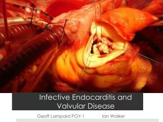
Nonpharmacological Treatment • Surgical removal, repair, and/ or replacement of infected valves or cardiac hardware • Support of vital functions
Pharmacological Treatment • β-Lactam antibiotics, such as penicillin G (or ceftriaxone), nafcillin, and ampicillin, remain the drugs of choice • The use of synergistic antimicrobial combinations may be required for certain pathogens to obtain a bactericidal effect • Once the infecting pathogen is identified, there are detailed guidelines for the treatment of each specific bacteria
Pathogen Specific Therapies (Slide 1 of 7) Native Valve Endocarditis caused by highly penicillin- susceptible (MIC≤ 0.12 mcg/mL) viridans group streptococci and Streptococcus gallolyticus (bovis)
Pathogen Specific Therapies (Slide 2 of 7) Native Valve Endocarditis caused by Streptococcus gallolyticus (bovis) and viridans group streptococci relatively resistant to penicillin (MIC> 0.12 mcg/mL)
Pathogen Specific Therapies (Slide 3 of 7) Prosthetic Valve Endocarditis caused by highly penicillin- susceptible (MIC≤ 0.12 mcg/mL) viridans group streptococci and Streptococcus gallolyticus (bovis)
Pathogen Specific Therapies (Slide 4 of 7) Prosthetic Valve Endocarditis caused by Streptococcus gallolyticus (bovis) and viridans group streptococci relatively resistant to penicillin (MIC> 0.12 mcg/mL)
Pathogen Specific Therapies (Slide 5 of 7) Native Valve Endocarditis caused by Staphylococci
Pathogen Specific Therapies (Slide 6 of 7) Prosthetic Valve Endocarditis caused by Staphylococci
Pathogen Specific Therapies (Slide 7 of 7) Prosthetic or Native Valve Endocarditis caused by Enterococci
Pathogen Specific Therapies Native and Prosthetic Valve Endocarditis caused by HACEKMicroorganisms
Endocarditis Culture Negative Therapies (Slide 1 of 2) • A patient with an acute clinical presentation of native valve infection should be started on antibiotic coverage for S aureus, β-hemolytic streptococci, and aerobic Gram negative bacilli • A patient with an subacute clinical presentation of native valve infection should be started on antibiotic coverage for S aureus, viridans group streptococci, HACEK, and enterococci
Overall Monitoring of Infective Endocarditis • Fever usually subsides within 1 week of initiating therapy • Echocardiography should be completed after completion of antibiotic therapy to establish a new baseline heart function • Blood cultures should be negative within a few days of starting antibiotic therapy
Patients at Highest Risk of Endocarditis • Prosthetic cardiac valve or prosthetic material used for cardiac valve repair • Previous infective endocarditis • Congenital heart disease (CHD) • Unrepaired cyanotic CHD, including palliative shunts and conduits • Completely repaired congenital heart defect with prosthetic material or device, whether placed by surgery or by catheter intervention, during the first 6 months after the procedure† • Repaired CHD with residual defects at the site or adjacent to the site of a prosthetic patch or prosthetic device (which inhibit endothelialization) • Cardiac transplantation recipients who develop cardiac valvulopathy
Summary Endocarditis typically presents as fever Antibiotic treatment durations differ significantly when treating native vs. prosthetic valve infections There are specific guidelines for each pathogen causing endocarditis Patients at the highest risk of infective endocarditis should receive prophylactic antibiotic therapy
References • Veverka A, Crouch MA, Odle BL. Chapter 89. Infective Endocarditis. In: DiPiro JT, Talbert RL, Yee GC, Matzke GR, Wells BG, Posey L. eds. Pharmacotherapy: A Pathophysiologic Approach, 9e. New York, NY: McGraw-Hill; 2014. • Baddour LM, Wilson WR, Bayer AS, et al. Infective endocarditis in adults: diagnosis, antimicrobial therapy, and management of complications: a scientific statement for healthcare professionals from the American Heart Association. Circulation. 2015; 132:1435–86. • Gerber MA, Baltimore RS, Eaton CB, et. Al. Prevention of rheumatic fever and diagnosis and treatment of acute Streptococcal pharyngitis: a scientific statement from the American Heart Association Rheumatic Fever, Endocarditis, and Kawasaki Disease Committee of the Council on Cardiovascular Disease in the Young, the Interdisciplinary Council on Functional Genomics and Translational Biology, and the Interdisciplinary Council on Quality of Care and Outcomes Research. Circulation. 2009;119:1541–15