Download
1 / 25
250 likes | 285 Vues
Learn about the natural history, communicable diseases, types of infection, ways of transmission, and microbial diseases in the oral cavity. Discover the oral manifestations of bacterial, viral, and other infections.

E N D
Course and forms of infection Special in oral cavity MUDr. Lenka Černohorská, Ph.D.
Natural history of infectious disease • The incubation period: time between acquisition of the organism/toxin and 1st symptoms • The prodromal period: non specific symptoms (fever, loss of apetite) • The acute specific illness: characteristic signs and symptoms • The recovery period: the patient returns to health
Course and forms of infection • Acute (running nose) – hours to days • Subacute (endocarditis) – weeks • Chronic (HIV) – years
Communicable diseases • endemic – low level in a specific population (Malaria in African countries) • epidemic – much more frequently than usual (epidemic influenza in the winter) • pandemic– worldwide distribution (HIV)
Course and forms - others • Inapparent (subclinical) infection – without symptoms, asymptomatic although infected • Chronic carriers – once infected, body can not completly eliminate the pathogen after recovery • Latent state – after period of a time reactivation and recurrence of symptoms may occur (primary infection of herpes reside in a latent state in ganglion, causing reccurent labial herpes).
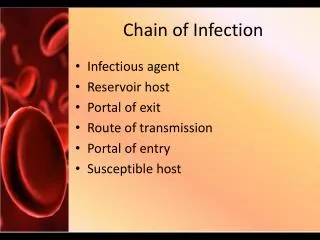
Types of infection • Exogenous origin – transmission from external sources • Endogenous – are caused by members of the normal flora (opportunist pathogen)
The ways of transmission • Inhalation – the airborne route (grip) • Ingestion – faecal contamination of food and water (salmonela) • Inoculation – sexual contact, needles, skin contact, blood transfusions or biting insects (HIV…)
Determinants of infection • Immune status (immunocompromited) • Genetic constitution • Age (children, old people) • Hormonal status • Pregnancy • Personal habits (smoking, drug using…)
Oral manifestation of bacterial infection • Scarlet fever- Streptococcus pyogenes, forming erytrogennoustoxin, exanthemaon soft palate curves and uvula, coalescent, scarlet red enanthema, white coated tongue, rich red with markedly exserted papillae –raspberrytongue, enlarged+painfullsubmandibular lymph nodes. • Skin rush – coalescent light red spots missing in perioral area • Therapy:penicillin Source: Wikipedia
Oral manifestation of bacterial infection • Diphtheria -Corynebacterium diphtheriae -pseudomembranous tonsilitis/laryngitis (croup). A thick, adherent pseudomembrane is present on tonsils or pharynx, may involve nasal mucosa, the pharyngeal wall andthe soft palate. Oedema involving the cervicallymph glands may occure in the anterior tissues of the neck – bullneck diphteria.
Oral manifestation of bacterial infection • Gonorrhoea- gonococci - tonsilitis and faryngitis
Oral manifestations of bacterial infection - Syphilis Primary –chancre – lips, tongue Secondary– mucous patches of the tonsils, soft palate, cheek Tertiary syphilis – gumma – hard palate, lips and tongue primary syphilis
Oral manifestations of bacterial infection - Syphilis Typical changes seen on permanent teeth (congenital) : • Hutchinson´s incisorsupper central incisors: barrel-shaped, crescentic notch at the incisal edge • Mulberry (moon) molarsfirst molar teeth have a roughened dirty, yellow, hypoplastic occlusal surface, poorly developed cusps resembling the surface of mullbery. mullberry molars Hutchinson´s incisors Source: kmil.trios.cz/ObrLues/hutchin1.JPG
TBC and Leprosy in oral cavity • TBC ulcers, granulomas, fissures, diffuse inflamatory lesions • Leprosy Tuberculoid leprosy - paraesthesia of the face, lips,tongue, palate, cheeks or gingiva Lepromatous leprosy – saliva is infectious, tooth loss, premaxillary bone resorption, saddle nose, intraoral nodules with tend to ulcerate
Viruses in oral cavity • Herpes simplex (HSV) HSV-1 (oral), HSV-2 (genital) Pathogenesis: Primary infection of children – innaparent or gingivostomatitis. Lesions on tongue, palate and gingiva + itching or burning sensation, blisters develope-enlarge-rupture- become encrusted. Gingiva – oedema, painfull, redish Latent infection – activation due to stress etc. - herpes labialis Dg. Smears from lesions to transport medium, antigen detection - immunofluorescence, serology Therapy: Acyclovir Dentist – frequent in paronychium, disease from work
Viruses in oral cavity • Varicella-zoster (VZV) Before the typical skin rash in chickenpox develops- lesions on the hard palate, pillars of the fauces and uvula: small ulcers surrounded by an area of erythema, quickly rapture Oral manifestations of zoster – severe pain like in toothache, one side enantem • Epstein-Barr (EBV) – infectious mononucleosis Petechial haemorrhages at the junction of the hard and soft palates – Holzel´s sign, later pseudomembrane tonsilitis, submandibular lymfadenitis. Edema of Waldeyer lymphatic circle cause problems with breathing
Viruses in oral cavity • Human herpesvirus 6 (HHV-6)- exanthema subitum/roseola infantum (6th exantematic disease). Virus is presentin the saliva, forms erythematous papules seen in soft palate anduvula (Nagayama´s spots) • Morbillivirus– in prodromal state, Koplik‘s spots on bucalmucose against molares – little white spots surrounded by dark red margine Koplik´s spots
Viruses in oral cavity Hand-foot-mouth disease • Coxsackievirus • Herpanginasmall (1-2 mm in diameter) papulovesicular lesions, with greysh-white surface surrounded by red areolae, especially in the palate/uvula - herpetic like - A 2, A 4-6, A 8 • Hand-foot-mouth disease– briht-red macules, in oral cavity later form oval/grey vesicles with red areolae http://pathmicro.med.sc.edu/virol/picorna.htm
HIV in oral cavity • Mycotic:Oral candidosis • Viruses: EB-virus specific damage - not painfull whitish corrugated lesions on the tongue margines – hairy leukoplakia, Kaposi´s sarcoma, herpetic gingivostomatitis and oral papilloma • Bacterial infection: gingivitis (linear marginal erytema/ ulcerous gingivitis), necrotizing stomatitis and necrotizing ulcerous parodontitis • Cervical lymphadenopaty, lymphomas
Mycotic infection • Oral candidiasis-Candida albicans Oportunne patogen. Predisposition – skin laesions, teethimplantates, malnutrition, diabetes mellitus, leukaemia, surgical operations, burnings, AIDS, immunosupresive therapy, antibiotic use • Therapy - local (clotrimazol), systemic (fluconazol, itraconazol, amphotericin B), autovaccines
Mycotic infection • Pseudomembranous candidiasis (thrush) – erythematous mucous membrane with milk white pablanes - newborns, older people, in immunocompromised is chronic, in HIV may spread into oesophagus • Erythematous (atrophic) candidiasis Acute - result after broad-spectrum antibiotic therapy, dysmicrobie of oral cavity – mucous membrane. Oral cavity: erythematous areas, burning sensation. Chronic - protetic stomatitis – frequently observed in elderly people wearing full dentures orthodontic appliances. Erythema+oedema of the mucosa. Therapy: removal of dentures at night, desinfection of dentures, carbohydrate diet • Hyperplastic candidiasis (candida leukoplakia)– chronic, from small, palpable, whitish areas to large, dense plaques, hard and rough to touch, on the inside surface of cheeks. High risk of malignant transformation! • Angular candidiasis-in one or both angles of the mouth, especially as a complication of protetic stomatitis, erythema and fissuring, vit. B12 deficiency
Salivary gland infection - bacterial • Acute suppurative parotitis(sialoadenitis suppurativa acuta) - painfull • Agens: alfa-haemolytic streptococci, S. aureus, haemophilus, eikenela, peptostreptococci • After intraabdominal operation acute afteroperation parotitis, first serous, later absceding
Salivary gland infection – viral - non suppurative • Parotitis epidemica(mumps) -replication in salivary duct epithelial cells – pain on chewing, redening of the opening of the duct, enlargement of gland + decrease of saliva secretion Complications: meningitis, orchitis of adult males (painfull+sterility), pancreatitis (increase of amylase level), nephritis Dg.: electron microscopy, serology (IgM detection – IF) • CMV- after reactivation of latent infection sialoadenitis (AIDS, immunosupression, cytostatic therapy) • HIV-minority - xerostomia and enlargement of the major salivary gland+ Sjögren´s syndrom (dry keratoconjunctivitis and progresive polyartritis)