Download
1 / 48
490 likes | 723 Vues
Care of the Respiratory Patient. Pneumonia Respiratory Failure. Function of the respiratory system. Exchange of gases between air and blood Exchange of gases between blood and cells Filter, warm, and humidify air Speech Sense of smell Maintenance of homeostasis.

E N D
Care of the Respiratory Patient Pneumonia Respiratory Failure
Function of the respiratory system • Exchange of gases between air and blood • Exchange of gases between blood and cells • Filter, warm, and humidify air • Speech • Sense of smell • Maintenance of homeostasis
Muscles required for breathing • Diaphragm • Intercostals • Abdominals • Neck, collarbone
Control of respiration • Center at base of brain (medulla oblongata) • Baroreceptors (carotid and aorta) • Both detect O2 and CO2 levels and cause adjustment in breathing rate
Other important sensors • Sensors in airways detect irritants and can • provoke a cough or • spasm as in asthma • Sensors in alveoli detect fluid build up and trigger rapid, shallow breathing • Sensors in joints and muscles detect movement and respond with increased rate and depth
Pneumonia • Acute inflammation of the lungs • Cause is infection • Initial diagnosis is by chest xray:
Causes, symptoms, treatment… Vary depending upon:
Community acquired pneumonia • Occurs in people with little-no contact with medical settings • Over 100 microorganisms can cause CAP • Streptococcus pneumoniae • Hemophilusinfluenzae • Enteric gram negative bacteria • Viruses • Pseudomonas aerugenosa • Viruses • Atypical organisms
Signs, symptoms of CAP • Malaise • Cough, usually productive • Dyspnea • Chest pain is pleuritic, adjacent to infected area • Can manifest as upper abdominal pain if site is close to the diaphragm • Signs include: fever, tachycardia, tachypnea, crackles, bronchial breath sounds
Diagnosis of CAP • Chest xray shows some infiltrate, but sometimes not until after 24-48 hours • Pulmonary embolism often mistaken for CAP if no sputum production • CBC and Chem Panel • 2 sets of blood cultures • Pulse oximetry or ABG • Usually no attempt to identify organism unless patient is critical! • ICU care required if mechanical ventilation required or if patient is hypotensive
Treatment of CAP • Antibiotics according to likely pathogen (“empiric treatment”) • 90% of patients with CAP treated empirically improve • Failure to improve suggests drug resistance • Most viral pneumonias resolve without specific treatment • Adults should have follow-up xray 6 weeks after treatment
Hospital acquired pneumonia • Develops at least 48 hours after hospitalization • Common causes are : • Gram negative bacilli • E. coli • Salmonella • Klebsiella • Proteus
Risk factors for HAP • Mechanical ventilation • Previous antibiotic treatment • Cardiac, pulmonary, hepatic, and renal comorbidities • Age > 70 • Abdominal or thoracic surgery • Dependent functional status
Signs, symptoms HAP • Same as CAP for nonintubated patients • In critically ill, mechanically ventilated patients: • Fever • Increased respiratory rate • Increased heart rate • Increased purulent secretions • Worsening hypoxemia
Diagnosis of HAP • Suspected on basis of new infiltrate • No symptom, sign, or xray finding is specific for the diagnosis • Aggressive testing (bronchoscopy, Gram stain with C&S, etc.) are controversial—are they accurate? Are the results colonization?
Treatment of HAP • Empirically chosen antibiotics • May begin with broad-spectrum (e.g., ceftazidime, imipenem, pipericillin/cipro, or gentamycin if no MDR risk) • Replaced with most specific drug after culture results obtained
LTC Facility acquired pneumonia • Usually gram negative bacilli, Staphylococcus aureus, Streptococcus pneumoniae, Haemophilusinfluenzae, and influenza
Risk factors for LTCFAP • Poor functional status • Impaired mood • Mental status decline • Swallowing impairment • Presence of tracheostomy
Signs, symptoms LTCFAP • Symptoms likely to be more subtle • Altered mental status • Anorexia • Weakness • Restlessness, agitation • Falling • Incontinence • Signs: ↓responsiveness, fever, tachycardia, tachypnea, wheezes, crackles, “wet breathing”
Diagnosis of LTCFAP • Xrays may be difficult in nursing home • Transfer to hospital for evaluation may be necessary • May start treatment without xray confirmation • May not have an infiltrate due to dehydration due to febrile pneumonia!
Treatment of LTCFAP • Should be hospitalized if 2 or more unstable vital signs • One dose of broad spectrum antibiotic before transfer
Pneumonia in the immunocompromised patient • Often caused by unusual pathogen • Diagnosis requires blood cultures, bronchoscopy, and chest xray • Overall mortality can be up to 20%
Preventative measures • Smoking prevention • Flu shot annually • Pneumonia vaccine (when?) • General health maintenance habits
Case finding • Be alert to signs and symptoms, particularly in the elderly • Perform through physical assessment • Provide patient education on CAP • Refer high risk patients to community follow up resources
Direct care • Monitor vital signs with SaO2 every 2 to 4 hours as indicated (may be hourly or more) • Monitor lung sounds as appropriate • TCDB • Antibiotics as prescribed • Maintain appropriate hydration • Maintain airway patency • Pain medication as indicated • Prevent additional sources of infection • Support patient’s mental and emotional status
Respiratory failure A syndrome in which the respiratory system fails in one or both of its gas exchange functions:
Acute vs chronic respiratory failure • Acute develops over minutes to hours • Chronic develops over days or longer, allowing time for renal compensation • pH decreased in acute, slightly decreased in chronic
Pathophysiology • Breathing is the process of moving air in and out of the lungs • Respiration involves three processes: • Transfer of oxygen across the alveolus • Transport of oxygen to the tissues • Removal of CO2 from the blood→ alveolus→ environment • Respiratory failure occurs from malfunctioning of any of these processes
Signs and symptoms • Dyspnea • Use of accessory muscles • Tachypnea • Tachycardia • Diaphoresis • Cyanosis • Altered consciousness • Eventual obtundation
Treatment respiratory failure • Acute failure requires ICU care • Correction of underlying cause • Supplemental oxygen • Control of secretions • Ventilatory support if necessary
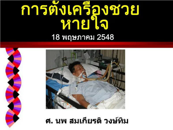
Mechanical ventilation • Required when the patient cannot maintain these volumes independently • If volumes are not maintained, oxygenation and dispelling of CO2 is compromised • Mechanical ventilation performs the functions that the lungs cannot, either temporarily or permanently
Ventilators • Wide range of ventilator sizes and complexities • Selection of ventilator depends on: • Desired result • Availability
What the ventilator provides… • There are many features, all of which cannot be covered here! • MAJOR components are:
Common ventilator modes:Assist control (AC) • Ventilator delivers a breath every time the patient initiates a breath • Ventilator breath will have either a preset tidal volume or preset peak pressure • There is usually a back-up breath rate that is set in the event the patient fails to breathe enough • Benefit: requires little work on the part of the patient
Common ventilator modes: Controlled mechanical (CMV) • Ventilator delivers breaths based on a preset rate regardless of the patient’s efforts • Not used if the patient is very alert and responsive
Synchronized intermittent mandatory ventilation (SIMV) • A preset breath based on pressure or volume is delivered regularly within so many seconds • Represents a “normal” respiratory rate for that patient • If patient initiates another spontaneous breath within that cycle, the ventilator will not deliver another mechanical breath so that the patient is not hyperventilated • Used when decreasing patient’s dependence on ventilator (“weaning”)
Continuous positive airway pressure (CPAP) • The patient initiates all breaths • The ventilator maintains a continuous level of pressure throughout the airways • Promotes oxygenation and decreases the workload of the heart and lungs
Positive end expiratory pressure (PEEP) • Similar to CPAP • Here the ventilator maintains a higher pressure in the lungs during the exhalation phase of breathing
Nursing responsibilities: mechanical ventilation • Validate accuracy of settings • Monitor patient response to O2 therapy • Collaborate with RT, MD, for appropriate delivery (mode, volume, O2 percent) • Maintain airway patency • Promote patient comfort • Monitor ABGs, SaO2 , and appropriate labs • Actively promote patient’s decreased dependence on mechanical ventilation if feasible