Tumor Markers
Tumor Markers. By: dr. hassan el- banna. Outlines: What is a Tumor Marker ? Characteristics of Tumor Markers Samples used for tumor markers measurement Tumor Markers Applications in Clinical Oncology Types of Tumor Marker Methodologies used for detection of tumor markers levels


Tumor Markers
E N D
Presentation Transcript
Tumor Markers By: dr. hassan el-banna
Outlines: • What is a Tumor Marker ? • Characteristics of Tumor Markers • Samples used for tumor markers measurement • Tumor Markers Applications in Clinical Oncology • Types of Tumor Marker • Methodologies used for detection of tumor markers levels • Frequency of Tumor marker estimation • Factors affecting Tumor Markers measurement
What is a Tumor Marker? The result of malignant transformation is malignant cells that, in each cycle of cell division, generate new malignant cells. In this process, the malignantly transformed cells acquire some new properties through which they differ from non-malignant cells of the same origin. The acquired properties can be either the changes in cellular morphology, physiology, or the changes in cell growth (behavior).Differences between normal and malignant cells are therefore being exploited in the detection of malignant cells (or malignancy in general), and the sub-stances that are being determined in this process are termed “tumor markers”.
Characteristics of an ideal Tumor Marker Ideal tumor marker shouldfulfill the following criteria: • High sensitivity Sensitivity means positive in disease. Tumor marker should give positive result in all patients. • High specificity Specificity means negative in health. Tumor marker should not present in healthy persons or present in small quantities. • Can be measured by a certain lab tool.
Organ specificity Tumor marker should be elevated specifically in certain organ. • Its level is correlated with prognosis Tumor marker should increase in bad prognosis and decrease in good prognosis. • Reliable predictive value Negative predictive value NPV:Proportion of subjects with a negative test result in patients who are correctly diagnosed. Positivepredictive value PPV:Proportion of subjects with a positive test result in healthy subjects. • Its level is directly proportional to tumor activity and/or size.
Tumor Markers Applications in Clinical Oncology • Screening and early detection of Cancer. • Differential diagnosis of malignancy. • Prognosis. • Monitoring disease progression/regression and determining the effectiveness of cancer treatment.
Types of Tumor Marker • According to sample type: • Cellular tumor markers: measured in tissues • Humoral tumor markers: measured in blood and other body fluids
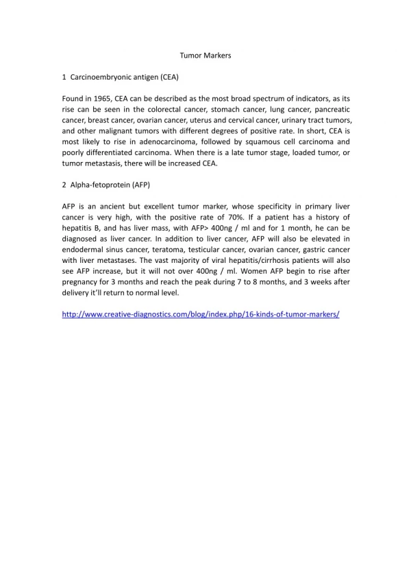
According to substance type: • Oncofetal Antigens: Oncofetal proteins are antigens that are normally produced during the embrional development. In adults, their production is limited or completely absent (stopped).Elevated concentrations in adults result from reactivation of certain genes that control cellular growth and are directly connected to malignant process. • Alfa-fetoprotein (α-FP) Liver and germ cell tumors (testis and ovary usedtogether with ß-hCG). • Carcino-embryonic antigen (CEA) Advanced adenocarcinomas, other GIT cancer, breast, liver and lung.
Cancer-associated antigens (CA): This is a heterogeneous group of more specific markers that comprises various membrane structures of tumor cells. • Cancer antigen-15.3 (CA-15.3) Metastatic breast cancer andadenocarcinomas. • Cancer antigen-19.9 (CA-19.9) Pancreatic, gastric and colon cancer • Cancer antigen-125 (CA-125) Epithelial ovarian cancer, advancedadenocarcinoma • Prostate-specific antigen (PSA)Prostate cancer.
Enzymes: Certain enzymes that are produced more in-tensely if a malignant process is occurring in the organism can also be used as tumor markers. • Prostatic acid phosphatase (PAP)Prostate cancer • Alkaline phosphatse (ALP)Liver pancreatic cancer • Lactate dehydrogenase (LDH)Lymphoma, seminoma, acute leukemia.
Special Serum Proteins: • ß-2microglobulin (ß-2M) • B-cell lymphoma multiple myeloma,chronic lymphocytic leukaemia • C-peptideInsulinoma • Ferritin Lung, liver, and prostatic cancer.
Hormones: • Malignant formations can alter the synthesis and secretion of various hormones. Quantitative and qualitative alterations of the synthesis and hormone secretion can therefore be the indicators of a malignant process and can be monitored as tumor markers. • Adrenocorticotrophic hormone (ACTH):Lung, prostate, gastrointestinal cancers, • Antidiuretic hormone (ADH):Small cell lung cancer • Beta-human chorionic (ß-hCG): Germ cell tumors (Choriocarcinoma) • Calcitonin:Breast cancer • Parathyroid hormone: (PTH) Breast cancer
Oncogens and Tumor suppressor genes: • Oncogenes: • Oncogenes (also called: proto-oncogenes) are normal cellular genes which on being activated found to be associated with cancer. They are involved in normal cellular processes such as differentiation and growth factors signaling. • C-myc • C-erbB2 • C-abl/bcr • N-myc
Tumor suppressor genes: • Tumor suppressor genes are normal genes that slow down cell division, repair DNA mistakes, or tell cells when to die (a process known as apoptosis or programmed cell death). When tumor suppressor genes don't work properly, cells can grow out of control, which can lead to cancer. • Retinoblastoma (Rb) • p53 • APC • BRCA-1, -2
Methodologies used for detection of tumor markers levels • Radioimmunoassay (RIA) • Colorimetric enzymes system “Enzyme-linked immunosorbent assay” (ELISA) • Spectrophotomertically • Chemiluminescence • Fluorescence
Frequency of Tumor marker estimation • Pre-therapy: to determine basal therapeutic level • During therapy: throughout the 1st 5 years • 1st – 2nd year (every 3 months) • 3rd – 5th year (every 6 months) • Before changing the therapeutic dose • When suspecting recurrence or metastasis • When there is a significant increase (every 2-4 weeks)
Factors affecting Tumor Markers measurement • In vivo factors • Levels of tumor markers may be affected by the following in vivo factors: • Renal failure and cholestatic disorders may increase some tumor markers levels. • Drug interactions may inhibit tumor markers levels. • Rheumatic disease may increase CA19.9 level • Rectal examination may increase PAP and PSA levels • Cigarette smoking may increase CEA level • Presence of inflammatory processes. • Presence of benign tumors and cirrhosis. • Some physiological conditions may interfere with tumor marker levels such as pregnancy (ßHCG, CA-125, CA-15.3, α-FP) and menstrual cycle (CA-125).
In vitro(lab)factors • Levels of tumor markers may be affected by the following in vitro (lab) factors: • Bad storage of serum samples • Hemolysis and icterus • Increased time of centrifugation • Contamination with other substances