Download
1 / 114
1.15k likes | 1.21k Vues
Learn about the anatomy and function of the cardiac system, including heart circulation events, heart sound occurrences, and extra heart sounds. Gain insights into murmurs and the cardiac cycle in this comprehensive guide.

E N D
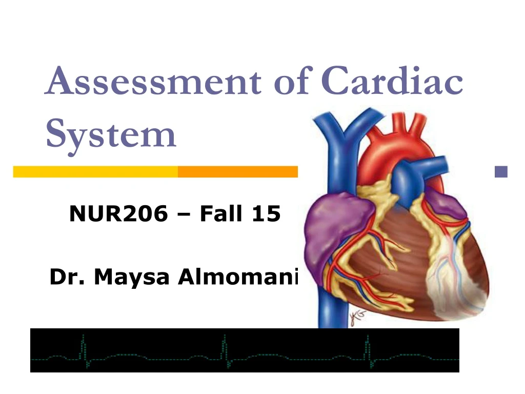
Assessment of Cardiac System NUR206 – Fall 15 Dr. Maysa Almomani
Pictures in brief • The right ventricle occupies the most of the anterior cardiac surface. • The inferior border of the right ventricle lies below the junction of the sternum and the xiphoid process. • The right ventricle narrows superiorly and meets the pulmonary artery at the base of the heart- the proximal surface of the heart at the right and left 2nd interspaces close to the sternum.
. • The left ventricle behind the right ventricle and to the left. It tapered inferior tip (apex) or PMI at the 5th interspace, 7 – 9 cm lateral to the midsternal line. It is approximate the size of quarter (1-2.5 cm in diameter). • Right atrium: form the right heart border while the left atrium is more superior and cannot be examined.
Great vessels with the Heart • Pulmonary artery: arise from the right ventricle • Aorta: from the left ventricle to the level of the sternal angle, where the arches backward to the left and then down. • On the right there is superior vena cava empties in the right atrium. Inferior vena cava empties blood from the lower part of the body.
Anatomy • Atrioventricular valves: Tricuspid and Bicuspid (Mitral) • Semilunar valves: aortic and pulmonic
. • The superior vena cava and inferior vena cava empties the unoxgenated blood into the RT atrium then it passes to the RT ventricle then to goes to the pulmonary artery then blood exchange happened in the lung and then return back through pulmonary veins to the LT atrium then to the LT ventricle then to the aortic arch to be distributed to the body.
Events in the cardiac cycle • The heart serves as a pump. • Systolic pressure: The period of ventricular contraction. The aortic valve open, the mitral valve close to prevent regurgitation back into the left atrium. • Diastole: Is the period of ventricular relaxation. Aortic valves close to prevent regurgitation of blood from aorta to left ventricle, the mitral valve open to allow bld flow from the left atrium to the relaxed LT ventricle. • During the systolic period the pressure of the ventricles rise to 120 mm Hg, and during diastolic pressure it falls to 5 mm Hg.
Left side Right side S1 during the systole: S2 during Dystole
How blood sounds occur? • During diastole: open of the mitral valves due to the increased pressure in the RT atrium slightly above that in the Lt ventricle. • During systole: the pressure in the LT ventricle rise so shutting the mitral and tricuspid valves to produce the S1 sound (Lub). • As ventricular pressure increase it quickly eject the blood into the aorta and forces the aortic valve to open. • When the LT vent eject most of its blood, ventricular pressure falls causing the aortic valveand pulmonic closer which produce the S2(Dub).
Splitting of the heart sounds • Same events occurs on the right side of the heart but the pressure on the LT side is more than that in the RT side. Also, events in the right happened later than those on the left.
Therefore: actually S1 (Lub): Caused by closure of Atriventricular valves (AV valves ; mitral & tricuspid) The start of systole Louder than S2 at the apex Heard with diaphragm Coincide with carotid artery pulse Coincide with the R wave of the QRS complex of the ECG Normal Heart Sounds
S2 (Dup) Closure of semilunar valves (aortic & pulmonic) Heard with diaphragm Loudest at the base Heard over pulmonic area (2nd left ICS) Normal splitting of S2 may occur with Inspiration (aortic & pulmonic valves closes separately) S1 S2 Inspiration A2 P2 Normal Heart Sounds
. • In children & young adults, a 3rd heart sound, S3 may arise from rapid deceleration of the column of blood against the ventricular wall. • In older adult, an S3, sometimes termed as S3 gallop, usually indicates a pathologic change in ventricular compliance. • although not often heard in normal adults, a 4th heart sound, S4, marks atrial contraction & indicates a pathologic change in ventricular compliance.
S3 & S4 occur during Diastolic Phase S3 (Ventriculargallop): A ventricular filling sound In early diastole after S2 Best heard at Apex or left lower sternal border. Best heard with diaphragm Low pitch Indicates decrease ventricular compliance Normal in children and young adults up to 40 yo, then considered abnormal Occur in patients with HF, aortic, mitral, or tricuspid regurgitation S4 (Atrial Gallop) A ventricular filling sound In late diastole immediately before S1 Best heard at the apex in left lateral position Best heard with the bell Soft low pitch Indicates decrease compliance of ventricle Could be normal in adults > 40 with NO evidence of cardiac disease Pathologic S4 occur with patients who have CAD, HTN, and aortic stenosis Extra Heart SoundsExtra slides for the instructor not for students
Cardiac Murmurs • Their sound is longer than heart sounds. • They attributed to the turbulent blood flow and may not indicate a pathological condition • In stenotic valve: ubnormal narrowing of valvular orifice obstruct the blood flow and cause the murmur like in aortic stenosis. • The valve may fail to close as in aortic regurgitation or insufficiency (regurgitant murmur).
Where to hear murmurs • Sound and murmur arise from mitral valve heard around the cardiac apex. • Tricuspid: the lower left sternal border. • Pulmonic valve heard best at 2nd and 3rd left ICS close to the sternum or higher or lower. • Aortic can be heard anywhere from the right 2nd to the apex
SA node Bundle of His AV node Conduction System in the Heart . • SA node (Sinus node) • Located in the RA • Acts as the cardiac pacemaker • Automatically discharge 60-100 times/min • AV node (atrioventricular) • Located at the atrial septum • Delay the impulse fired by the SA node before the impulse passes to the bundle of His • Muscular contraction follows; first atria, then ventricles • Muscular contraction produce electrical activities, resulting in a series of waves on the ECG. • P-wave: atrial depolarization • QRS complex: ventricular depolarization • T-wave ventricular repolarization or recovery
Definitions • Preload: the load that stretches the cardiac muscle prior to contraction; it is the volume of blood in the right ventricle at the end of diastole. • Afterload: the vascular resistant against which the ventricle must contract. • Stroke volume (SV): the volume of blood ejected with each heart beat. • Cardiac output: the volume of blood ejected from each ventricle during 1 minute. It is the product of HR and SV. • Stenosis: abnormally narrow valvular orifice that obstruct blood flow • Regurgitation: insufficiency of valvular orificethat allow blood to leak backward and produce a regurgitate murmur.
JUGULAR VENOUS PRESSURE • Systematic venous pressure is lower than arterial pressure. • Venous pressure depends on the ventricular contraction, but the walls of the veins contain less smooth muscle than walls of the arteries which reduce the venous tone and makes veins more distensible
JUGULAR VENOUS PRESSURE • The venous pressure changes are reflected in the height of venous column of blood in internal jugular veins known as jugular venous pressure JVP. • Pressure in the jugular vein reflects right arterial pressure which gives an indication of the function of the right heart homodynamic
. • JVP is best estimated from the internal jugular vein, usually the right since it has more direct anatomic channel into right atrium. • The internal lies deep in the sternomastoid muscle. The pulsation of the internal JV transmitted to the surface of the nick • If pulsation from the internal jugular cannot be identified then the external one can be used.
. • The JVP is measured in vertical distance above the sternal angle (bony ridge adjacent to the second rib). • The sternal angle is 5 cm above the right atrium regardless of the patient’s position. • JVP more than 4 above the sternal angle or more than 9 cm above the right atrium is considered high. • Technique for measurement will follow.
Health History • Chest pain: ask patients about history of angina pectoris, MI. • Dyspnea, orthopnea, (paradoxical noctural dyspnea (PND) • Cough • Fatigue • Cyanosis or pallor • Edema/swelling • Past cardiac surgery • Family cardiac Hx • Personal habits (risk factors)
. • Your basic Question: do you have pain or discomfort in your chest? • Is the pain related to exertion? • What kinds of activities bring on the pain? • How intense is the pain? • Does it radiate to: neck, shoulder, back, arm? • Associated symptoms like SOB, sweating, palpitation, nausea? • Does it wake you up At night? • what makes it better or worse
Terminology • Palpitation: unpleasant awareness of the Heartbeat. • Terms pt use like: skipping, racing, stopping, pounding. It does not necessary indicate heart disease • Ask questions like: are you ever aware of your heart beat? What it is like? Was it fast or slow? • Did they stop suddenly or gradually?
Shortness of Breath: uncomfortable awareness of breathing that is inadequate for the level of excursion. • It may represent heart or pulmonic problemand associated with orthopnea, Paroxysmal nocturnal dyspnea.
. • Orthopnea: dyspnea that occurs when patient is lying down and improves when patient is sitting up. • Paroxysmal nocturnal dyspnea: episodic of sudden dyspnea and orthopnea that awaken the patient from sleep, mainly after 1-2 hours after going to bed. Can be associated with cough and wheezes. • Edema: accumulation of fluid in the interstitial space.
Cholesterol Level: LDL <100 mg/dL HDL 40-60 mg/dL Triglyceride < 200 mg/dL Total Cholesterol <200 mg/dL Risk factors (“risk equivalents”: Smoking HTN HDL < 40 Family Hx. Age (men >45, women > 55) DM Atherosclerotic Diseases Desired goal for LDL level depends on # of risk factors: <160 if 0-1 risk factors <130 if 2+ or multiple risk factors <100 if CHD Health Promotion Behaviorsnot for students
Dietary management Low saturated fat cream, cheese, ice-cream, bacon, butter, chocolate) Low cholesterol High fiber Weight reduction Assess BMI Overweight BMI > 25 Obese, BMI >30 Regular exercise Aerobic exercise Regular 20-60 x 3-5 times/week Screening for HTN Health Promotion Behaviorsnot for students
Not for students • Drug therapy should be started with stage 1 hypertension 140/159 or diastolic BP 90 to 99 mm Hg • 55 years have %90 lifetime risk for developing HTN • More than 1 of every 2 older adults than 60 has HTN. • The relationship between HTN and heart disease is continuous
Blood pressure indicates cardiac function • Risks for HTN include: • low physical activity. • Family history • Increase sodium. • Low potassium. • Alcohol consumption
;Health promotion and counselling • Preventing hypertension • Preventing cardiovascular disease and stork • Lowering cholesterol and low density lipoprotein (LDL) • Lifestyle modification and risk intervention, including healthily eating and counselling about weight and exercise. (page 347)
;Preventing hypertension • Screening of adults 18 years and older for high blood pressure • Normal 120/80 • Pre-hypertension S: 120-139 and D: 80-89 are pre-hypertension. • stage 1 hypertension 140/159 or diastolic BP 90 to 99 mm Hg • Screening of risk factors: • Physical inactivity • Family history • Excessive intake of sodium , insufficient intake of K • Alcohol consumption.
;; • Preventing cardiovascular disease and stork: • Screening of adults at age 20 and 40 years (page 342) • Lowering cholesterol and low density lipoprotein (LDL) (known as "bad" cholesterol).
Techniques for Examination • First step is to assess the BP after: • setting the patient for 5 min, • choose the correct size cuff, • make sure the bladder is centered above the brachial artery, • inflate the cuff about 30 mm Hg above the pressure at which the brachial or radial pulse disappear.
Techniques for examination • The Neck Vessels • Jugular vein • Inspection • JVP • Carotid artery • Palpation • Auscultation
Jugular Venous Pressure (JVP) • Reflects right atrial pressure • The height of the venous column of blood in the internal jugular veins. • pulsation of internal jugular vein transmits to the surface of the neck. • pulsations of the external jugular vein is less reliable. • Distinguish internal jugular pulsation from those of carotid artery (page 350).
JVP Measurement: Notes on JVP measurement • Difficult in children and younger than 12 years old, so it is not useful to measure it. • Turn the patient head away from the side you are inspecting. • Use tangential light to see both sides of the neck. • As starting point, degree Raise the head of the bed to about 30 • Modify the bed to the right position for the patient.
Jugular Venous Pressure JVP • Hypovolemic patients may need to lie flat before you see the veins. • When JVP increases, the patient may need to be elevated 60-90 degrees. • Increased JVP may suggest Rt side HF, tricuspid stenosis or superior vena cava obstruction. • In patients with COPD, veins collapsed with inspiration and pressure elevated during expiration. • Unilateral distension of jugular vein may be due to local obstruction or kinking.
. • To estimate the value of the JVP • Find the highest point of oscillation in the internal jugular vein • If you are not able to see the pulsation of IJV look for them in the Ext JV. Use the point above which the external veins appears to collapse. • Measure the vertical distance of the pulsation from the sternal angel (bony ridge adjacent to the second rib) and add 5 cm to the distance • Normal JVP is < = 4 cm above the sternal angle or < = 9 cm above the right atrium.
Carotid Artery • Provide information about cardiac function and detect stenosis or insufficiencyof the aortic valve. • Assess amplitude (small, thready, or weak; bounding) and contour: thespeed of the upstroke, theduration of its summit and the spead of the downstroke (brisk; smooth, round, follows S1 immediately, rapid). • Patient should be lying in 30 degree. • Place your index and middle finger or thump in the lower third of the neck, press posteriorly and feel for pulse.