Download
1 / 25
350 likes | 867 Vues
Total Knee Arthroplasty in Valgus knee. H.Makhmalbaf MD Consultant Knee surgeon Mashad University. The Typical patient. Sever valgus deformity Elderly woman in7th or8th decade Has had “knock knees” Also had patellofemoral problems Valgus deformity progresses Loss of lateral joint space

E N D
Total Knee Arthroplasty in Valgus knee H.Makhmalbaf MD Consultant Knee surgeon Mashad University
The Typical patient • Sever valgus deformity • Elderly woman in7th or8th decade • Has had “knock knees” • Also had patellofemoral problems • Valgus deformity progresses • Loss of lateral joint space • Attenuation of MCL
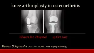
The typical patient • PFJ involvement & patella dysplasia on X-Ray • Patella subluxation & concave patella • Patella alta & a very thin patella • Chondrocalcinosis & calcification of menisci • Long leg film of tibia shows valgus bow
The typical patient • The valgus tibia is problematic if using intramedullary guide • The long leg film is necessary to template • The diagnosis is usually OA or RA • Hypermobile knee or hyperextention • In RA might have stiff knee & flexion cont.
Clinical features of valgus vs varus • Valgus knees tolerate significant damage • In windblown knees varus knee is more symptomatic • Varus knee should be operated on 1st • Source of deformity is also different • Medial tibial plateau is deficient & tibial joint line in sever varus in varus knees • The femoral joint line is still in 5-7 valgus
Valgus vs varus knee • In valgus knees, is from the femur • The tibial joint line usually in neutral • The femoral joint line is in marked valgus • Deformity due to hypoplasia of lat.femoral condyle • Both distally & posteriorly • The tibial defect in valgus is contained
Variants of valgus • Based on degree of deformity • Status of MCL, osseous deficiency & • The amount of release must be performed • Variant I: mild & correctable deformity • Variant II: greater deformity ,intact soft tissue • Variant III: stretched MCL, Marked bone loss & deformity & lateral contracture
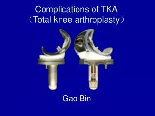
Type of prosthesis • In variant I: P CR or PS may be used • In variant II: may require a PS implant • Invariant III: a PSC (constrained) should be used • Complete set of instruments if preferred
Lateral femoral condyle hypoplasia • Is present in sever valgus deformity • Exists both distally & posteriorly • In distal femoral resection don’t cut to the defect • The deficiency must be augmented • If not, causes large extension gap • And elevation of joint line • And mid-flexion laxity, distorting kinematic
The angle of DF cut • 5 degree is preferred • 7 degree cut is better for balance but? • 5o or less to overcorrect the deformity • Less tension on MCL on WB • But needs more lateral release • Distal metaphyseal bow • More medial point of entrance in the notch
Balancing the knee in flex.& extension • Create symmetric flexion extension gaps • Valgus knees could be balanced in flexion & extension independently • In flexion the tissues are not tight • Nor the medial tissues are lax in flexion • Draw Whiteside line & transepicondylar ax • In sever valgus int rotation of femoral component is necessary
Flexion extension balance • Lateral release, after bone cuts • Put spacers, varus & valgus tests • Inverted cruciform release • LCL, poplliteal, & Biceps tendon release • PS in sever valgus & CR in mild one • Surgical approach
Summary • Associated with PFJ disease • Lat femoral condyle hypoplasia • Medial laxity, a valgus tibial bow • Conservative bone cuts • Cruciform release of ITB & capsule • Protect peroneal nerve • PS or CR knee