
Supplementary Figure 3: Axonal swellings in injured spinal cord from YFP-H mice. (A-D) Fluorescence composites showing spread of axonal dystrophy along dorsal column (DC) fibers from YFP-H and Wld S /YFP-H mice at 10 h (A) and

E N D
Presentation Transcript
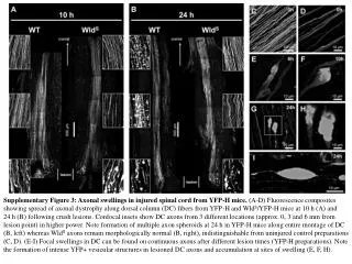
Supplementary Figure 3: Axonal swellings in injured spinal cord from YFP-H mice. (A-D) Fluorescence composites showing spread of axonal dystrophy along dorsal column (DC) fibers from YFP-H and WldS/YFP-H mice at 10 h (A) and 24 h (B) following crush lesions. Confocal insets show DC axons from 3 different locations (approx. 0, 3 and 6 mm from lesion point) in higher power. Note formation of multiple axon spheroids at 24 h in YFP-H mice along entire montage of DC (B, left) whereas WldS axons remain morphologically normal (B, right), indistinguishable from uninjured control preparations (C, D). (E-I) Focal swellings in DC can be found on continuous axons after different lesion times (YFP-H preparations). Note the formation of intense YFP+ vesicular structures in lesioned DC axons and accumulation at sites of swelling (E, F, H).