Download
1 / 51
510 likes | 752 Vues
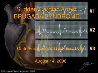
Cardiac Emergencies: What is My Role?. Kate Jessop RN, BSN Valley Hospital Medical Center Las Vegas, Nevada. ELECTRICAL CARDIAC ISSUES: ARRHYTHMIAS. Cardiac Properties Influencing Output. Automaticity – ability to spontaneously repolarize

E N D
Cardiac Emergencies: What is My Role? Kate Jessop RN, BSN Valley Hospital Medical Center Las Vegas, Nevada
Cardiac Properties Influencing Output • Automaticity – ability to spontaneously repolarize • Conductivity – ability include all cells of muscle mass once impulse initiated. • Contractility – ability to sustain contraction and empty • Rhythmicity – ability to spontaneously depolarize at regular intervals • Excitability – ionic exchange across cell membranes that facilitates electrical activity
Normal Cardiac Rhythm Determinants • SA node • AV node • Bundle of His • Bundle branches • Purkinje fibers
Cardiac Electricity • 1 - Sinoatrial Node (SA) node - Starts or Generates Impulse • 2 - Atrioventricular Node (AV) node - Filters Impulse and Protects Ventricle • 3 - Bundle of His • 4- 6 - Electrical Conduction of the Ventricles • 7, 8 and 9 - Left Ventricle, Septum and Right Ventricle • 10 - Right Bundle Branch
Rhythms Classified as Life Threatening • Extreme bradycardia • Asystole/ Ventricular standstill • Ventricular tachycardia • Ventricular fibrillation • Pacer dependency with loss of capture • Pulseless electrical activity (PEA)
Nursing Considerations • Place patient on cardiac monitor. • Provide oxygen if available. • Think about intravenous access – assist MD if needed. • Determine if patient is stable or unstable with MD. • Provide comfort to the patient. • Understand medications that are being given, adenocard / cardizem. • Need for cardiology consultation if needed. • Aspirin therapy if indicated.
Fast Heart Rates- Tachycardias • Sinus Tachycardia - Heart rate originating from the sinus or SA node with a rate greater than 100 beats per minute (BPM).
Causes of Sinus Tachycardia • Fever • Anxiety • Malignant Hyperthermia • Hypovolemia - Fluid or Blood Loss • Drugs - Cocaine, Nicotine, Caffeine, Amphetamines • Sepsis - Shock • Heart Failure • Anemia • Drug Withdrawal • Hypoxia- Low Oxygenation • Numerous Other Causes……
Treatment of Sinus Tachycardias • Treat underlying problem - Dehydration, fever, drug use, blood or fluid loss. • Intravenous fluids - Normal Saline or blood if needed for anemia. • Oxygen and rest. • Treat medical problem with physician consult or order. Consider heart failure, sepsis or pulmonary emboli.
Supraventricular Tachycardia • Heart rate originating from above the atrioventricular (AV) node
Signs and Symptoms of Supraventricular Tachycardia • Pounding sensation in chest. Rapid beating of heart - Racing • Chest pain • Shortness of Breath - Dyspnea • Fast breathing – Tachypnea • Dizziness or light headedness • Diaphoresis or sweating • Anxious
Problems with Supraventricular Tachycardias • Decrease in Cardiac Output - Inadequate amount of blood being pumped out of the heart as a result of the heart not having enough time to adequately fill the ventricles. • Cardiac muscle itself becomes tired and does not get adequately oxygenated blood to the myocardium. • Heart rate is usually above 150 and below 200 beats per minute (BPM).
Treatment of Supraventricular Tachycardias • Vagal Maneuver’s – Bear down, hold their breath and push down. • Ice water • Adenocard /Adenosine - Given rapid intravenous (IV) push followed by rapid fluid infusion as medication has a rapid half - life. • Diagnose underlying condition- Atrial arrhythmias such as atrial fibrillation or flutter. • Cardizem or calcium channel blocker intravenous under MD order.
Atrial Fibrillation • Irregularly irregular heart beat • Can cause blood clots • Loss of “atrial kick” • Greatly increases risk of stroke
Atrial Fibrillation-Diagnosis • Electrocardiogram readings • No definitive P waves • No cardiac equipment available? • Palpate a pulse • Beat will be irregularly irregular
Paroxysmal Atrial Fibrillation • Atrial fibrillation that starts suddenly and resolves suddenly without medical treatment • Can still cause stroke • Patients may experience dizziness, rapid heart rate, shortness of breath and palpitations • “Holiday heart”—sensitivity to alcohol causing atrial fibrillation • Often from large quantities of alcohol • Occasionally patients may have a high sensitivity to alcohol—just one drink can cause the arrhythmia
Ventricular Tachycardia • May or may not have a pulse associated with rhythm • IV Amiodarone or lidocaine can be given • With no pulse treat as ventricular fibrillation and defibrillate immediately • If no pulse begin cardiopulmonary resuscitation after defibrillation
Ventricular Fibrillation • Disorganized and chaotic heart rhythm with no pulses present. • Treatment immediate unsynchronized defibrillation • IV epinephrine
Complete Heart Block • Atria does not communicate with the lower ventricles • Treatment includes transcutaneous pacing or insertion of transvenous pacemaker
Asystole • Cardiac Standstill – No cardiac activity • Begin compressions and advanced cardiac life support (cardiopulmonary resuscitation and medications) • IV medications to include epinephrine and atropine
Myocardial Infarction • Decrease or stop in blood flow to the myocardial tissue causing cell death • Can be measured by troponin and CK-MB laboratories • Watch for ST changes on the electrocardiogram • Interventions: • Place patient on cardiac monitor • Oxygen • Aspirin (324 mg chew) • Nitroglycerin • Morphine
Diabetes and Myocardial Infarction • Diabetic patients often have neuropathy and may not feel the pain of a myocardial infarction (heart attack) • Atypical presentations are more common for diabetics; may include: • Nausea and/or vomiting • Shortness of breath • Cough • Wheezing • Anxiety • Diaphoresis
Pericarditis • Inflammation of the outer lining of the heart • Primary symptom: chest pain • Pleuritic in nature • Often relieved by sitting up, leaning forward, and taking shallow breaths • Other possible symptoms: • Tachycardia • Low grade fever • Malaise • Pericardial friction rub • Treatment: • Aspirin/Ibuprofen • Steroids
Pericarditis • EKG • Diffuse ST elevation with PR segment depression
Myocarditis • Inflammation of the ‘muscle’ of the heart • Symptoms: • Fatigue, dyspnea, palpitations, precordial discomfort, slight rise in serum enzymes • Endomyocardial biopsy is needed for definitive diagnosis • Treatment: • Supportive therapy • No competitive sports for at least six months
Endocarditis • Inflammation of the inner lining of the heart, including the valves • Bacterial, viral or fungal infection • At-risk populations: • Children with congenital heart disease, adults with mitral valve prolapse, rheumatic heart disease, intravenous drug abusers, and patients with prosthetic valves • Diagnosis: • Malaise, loss of appetite, fatigue, weight loss, night sweats, fever, new or changed heart murmur, positive blood cultures • Treatment: • Antibiotics—first broad spectrum and the based on blood culture results, usually long term treatment is needed • Emergent surgical intervention if congestive heart failure occurs due to valve dysfunction
Cardiac Arrest / Code Blue • What is cardiac arrest?? • Who responds?? • What is each persons/ providers role in the event?? • Role of the team leader • Roles for team members: Airway, Compressions, Intravenous access, Medications, Electrocardiogram, Recording and talking with family • Termination of efforts
What is Cardiac Arrest/Code Blue?? • Cardiac arrest is the abrupt change in the patient’s medical health, through respiratory or cardiac failure, by either mechanical or chemical imbalances. • What causes cardiac arrest?? Numerous and varied events such as myocardial infarction - MI, respiratory arrest, pneumothorax, metabolic entropy, trauma and numerous others not listed.
Recognition and Action • First and foremost, is the nurses ability to recognize that the patient’s condition has rapidly deteriorated. • Ensure that the patient is indeed connected to all appropriate monitoring devices and that they are functioning appropriately (i.e. monitor is plugged in or batteries are charged, oxygen is connected, etc.). • Establish unresponsiveness and call for MD and additional team members for assistance.
Your Roles in an Arrest or Code Blue • There are numerous important roles during a cardiac arrest. As primary nurse you should be the most familiar with the patient and have useful information for the MD. • In some countries, nurses can function as team leader until relieved by physician. If not, the primary nurse should establish unresponsiveness, lack of pulses and delegate someone to begin compressions along with getting a nurse or respiratory therapist to begin rescue breathing until advanced assistance arrives.
RN or MD as Team Leader • Either a RN or MD may be a team leader depending on hospital or governing rules. From a nursing standpoint, the nurse should be not only informative as to what transpired but also provide insight as to what may have caused the initial arrest. • Secondly, the nurse can provide important medical history and surgical information to the team leader, whomever it may be.
Compressions • Compressions in the event of cardiac arrest are equally as important as ventilation - the blood and medications need to be circulated whether by a pulse or artificial compressions. • One nurse or technician needs to be assigned to compressions with an alternate - the primary provider will get tired and need to be rotated. This will ensure adequate compressions and maximize perfusion to the brain and other vital organs.
Compressions (Cont.) • Compressions need to be performed at least 100 times per minute and to a depth of 5 centimeters. • Compression should be stopped only when absolutely necessary. • If two rescuers are present, the rate for two rescuer CPR is 30 compressions to 2 breaths. • It is the goal of CPR is to establish the return spontaneous circulation (ROSC).
Oxygenation and Respiratory Care • Airway and breathing are of paramount importance. If a respiratory therapist is not present or available, then one nurse should be assigned to manage the airway using the highest level of training present - whether that be bag valve mask (BVM) or advanced placement of an endotracheal tube (ET). • Ensure that suction is available as the patient may vomit or have excessive secretions that need to be removed in order to increase adequate oxygenation efforts.
Medications • Whether the team leader is the physician (most cases), or an RN leads until the MD arrives, it is important to know what medications to administer as well as what routes they may be given. • Some medications can be given down the endotracheal tube until an IV can be established. Most medications are given IV, but an interosseous line (one drilled into the bone) is also fitting for administration of medications, including blood products.
Recording • One nurse should be solely assigned to keep record of all events that are transpiring. It is the responsibility of this nurse to inform the team leader of the number of doses of each medication that has been given, as well as the time interval that has elapsed since that medication was last used. • Additionally, the nurse should also annotate when CPR is ongoing or when there is a change in the patient’s rhythm or condition.
Recording (Cont.) • Additional observations include when defibrillation or transcutaneous pacing is used, and the results of either of these interventions. Endotracheal tube size as well as depth is also of importance to the record. • Lastly, termination of the event must also be recorded to include providers present, as well as team leaders signature and actual time of death.
Successful Resuscitation • If the event is successfully survived, whether it be a simple case of a patient requiring an advanced airway or an additional medication needing to be given to reverse a narcotic, the nurse must still communicate with the physician to ensure where the patient needs to be moved or transferred to, and what was the underlying cause of the potentially life altering event. This may include transferring to an intensive care unit or possibly a higher level of care facility.
Patient Demise • In the event that the patient’s condition deteriorates and despite numerous attempts at returning circulation or spontaneous breathing and the actions are now proving futile, the arrest must be called. • Considerations must be given to quality of life post arrest as well as potential brain injury from the event, also known as an anoxic brain event.
Termination of Efforts • When the decision to terminate efforts has been decided upon, it is necessary to talk with the patient’s primary MD as well as family members. • When possible, the family should be notified in person or may be present during the resuscitation. The team leader needs to talk with the family and give them frequent updates in order for them to feel like everything possible is being done for their loved one.
Aftercare • Once the arrest is completed, the patient and room need to be cleaned so that family may come in and be at the bedside. • It is the responsibility of the nurse and MD to ensure all needles and syringes are disposed of properly and that the patient is presentable -meaning that blood, body fluids and refuse has been picked up and that there are no dangers to anyone entering the room.
Aftercare (Cont.) • Depending on the location of the arrest and surrounding circumstances an autopsy or investigation may be required. Consult the administration as well as hospital policies in your facility for direction. • Some patients may have requests for cremation or burial. Honor those requests as well as the wishes of the family. If organ donation is possible notify the appropriate agencies and prepare the body accordingly.
Team Leader Responsibilities • Evaluation of the event (i.e. what was great, what needed work or what was missing). • Completion of documentation. • Discuss occurrence with primary MD as well as family. • Thank members of your team for their hard work. Answer questions that the staff may have in a respectful manner and be supportive to responses. Give constructive praise and encouragement.