Download
1 / 63
670 likes | 797 Vues
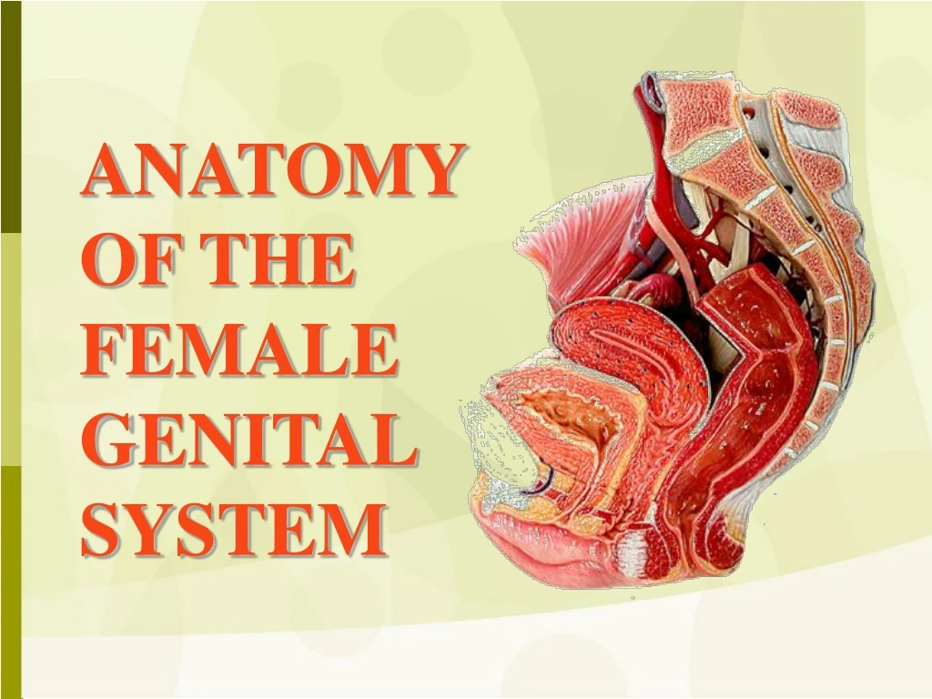
Dive into the intricate details of the female genital system, from the vulva to the vagina, exploring structures, functions, and anatomical relations. Discover key elements such as the mons veneris, Clitoris, Labia Majora and Minora, and more.

E N D
THE VULVA • 1. Mons Veneris: • a pad of fat overlying the symphysis pubis and covered by skin & hairs. • 2. Clitoris: • an erectile cavernous structure below the symphysis pubis. • formed of a small glans and two corpora cavernosa.
THE VULVA • 3. Labia Majora: • he outer 2 skin folds, raised by underlying fat, and passing back from the mons veneris to the perineum. The outer skin is covered by hairs while the inner medial surface is smooth, hairless and contains sebaceous and sweat glands. • 4. Labia Minora: • 2 thin folds of modified skin situated medial to the labia majora.
THE VULVA • 5. The Hymen: • a membrane, situated about 2 cm from the vestibule that demarcates the external from the internal genital organs, and partially closes the vaginal orifice.
THE VULVA • 6. Bartholin Glands: (Greater Vestibular Glands): • bilateral compound racemose glands • secrete mucus during sexual excitement • situated deep in the labia majora, at the junction of the posterior and the middle thirds • Its duct is 2 cm long and opens between the hymen and the labium minus. • 7. Vestibule: • the area between the inner aspects of the labia minora and the fourchette. • Structures that open in the vestibule are: • Urethra • The Bartholin glands ducts. • 3. The vagina. • 8. Vestibular bulbs: • oblong masses of erectile tissue that lie on each side of the vaginal introitus • 9. External urethral meatus: • a triangular slit in the anterior part of the vestibule below the clitoris in which the urethra opens. • 10. Skene’s duct: • 2 blindly ending para-urethral tubules which open in the floor of the urethra, few millimetres form the external urethral meatus.
Types of hymen Bi-perforate imperforate Virgin Deflorated Cribriform
Blood Supply • Arterial Supply: • Internal pudendal artery: The terminal branch of the anterior division of the internal iliac artery that ends as the dorsal artery of the clitoris. • Branches from the femoral artery, supply the anterior part. • Superficial and deep external pudendal arteries. • Venous Drainage: • The veins draining the vulva form a venous plexus from which veins accompany their corresponding arteries. The veins draining the clitoris join vaginal and vesical venous plexuses.
Lymphatic drainage and Nerve Supply • Lymphatic Drainage of the vulva • From the skin and appendages, to the superficial inguinal lymph nodes, to the deep inguinal and femoral lymph nodes of which the lymph node of Cloquet drains the clitoris directly. • From the former superficial group, lymphatic channels pass to the deep pelvic nodes including; the external iliac, common iliac, then para-aortic lymph nodes. • Nerve Supply of the vulva • The vulva is supplied mainly from the pudendal nerve (S 2, 3 & 4). • Additional sensory nerves are supplied from; the Ilio-inguinal nerve (L1), the genital branch of genito-femoral nerve (L 1,2), and the posterior cutaneous nerve of the thigh.
THE VAGINA • A fibromuscular tube from the vulva to the uterus forming an angle of 60° with the horizontal plane. • Length: • anterior wall is 8-9 cm • posterior wall is 10 -11 cm • Vaginal Fornices: • The cervix projects in the upper blind end of the vagina that forms a pouch (vaginal pouch) around the cervix and is divided into four fornices : two lateral, anterior and posterior (deeper) fornices.
Anatomical Relations of the Vagina • Anteriorly: • Upper 1/3: trigone of urinary bladder • Lower 2/3: urethra. • Posteriorly: • Upper 1/3: peritoneum of Douglas pouch. • Middle 1/3: ampulla of rectum. • Lower 1/3: the perineal body. • Laterally: • Lower end: Bulbocavernosus muscle, vestibular bulb, and Bartholin gland. • 1 cm above orifice: urogenital diaphragm • 2½ cm above the orifice: levator ani muscle with the pelvic fascia above it. • The lateral fornix gives attachment, to the lower part of the cardinal ligaments. • The ureters pass through the cardinal ligaments 1 cm lateral to the vagina.
Vaginal Supports • Ligaments attached to the upper vagina: • Pubocervical ligament anteriorly, • Mackenrodt’s ligament laterally, • Uterosacral ligament posteriorly. • Levator ani muscles: pubo-vaginalis part • Triangular ligament, and the Perineal membrane. • Vaginal fascia: Connective tissue fascia that condenses anteriorly forming the vesico-vaginal fascia and posteriorly forming the recto-vaginal fascia.
Histology of the Vagina • The cut section of the vagina is “H” shaped with approximation of the anterior to the posterior vaginal walls. It is formed of • Three layers; • mucosa, formed of squamous epithelium without glands, the • musculosa, which is fibromuscular with some fibres from the levator ani inserted into it, and the • adventitia, which is connective tissue continuous with the paracolpos.
Blood Supply • Arterial supply: • The vaginal artery (from internal iliac artery) • Additional branches from: • Middle rectal artery (from internal iliac artery) • Inferior rectal artery (from the internal pudendal artery, of the internal iliac artery) • Venous drainage: • A plexus around the vagina (the vaginal plexus), drain into the internal iliac vein by veins that accompany their corresponding arteries.
Lymphatic drainage and Nerve Supply • Lymphatic drainage of the vagina • lower 1/3 drains to the inguinal lymph nodes, • upper 1/3 follows lymphatic drainage of the cervix, • middle 1/3, drains in both upper and lower directions. • Nerve supply of the vagina: • The pudendal nerve gives sensory fibres to the lower vagina.
Applied Anatomy • Vaginal Prolapse: Weakness of the vaginal supports (ligaments, fascia and muscles) may lead to: • descent of anterior vaginal wall (cystocele or urethrocele), • descent of posterior vaginal wall (rectocele or enterocele), or • descent of the vaginal vault after hysterectomy (vault prolapse).
Applied Anatomy • The posterior fornix: • offers a passage to the pouch of Douglas for performing culdoscopy, culdocentesis and for drainage of a pelvic abscess. • The lateral fornix: • The ureter lies 1-2 cm lateral to it so that it may be injured during clamping the angle of the vagina in hysterectomy operation.
Applied Anatomy • Pudendal nerve block: • Transvaginal injection of a local anaesthetic solution around the pudendal nerve as it passes around the ischial spine gives a local anaesthesia sufficient for minor operations on the vulva and vagina, and has been used for low forceps operations in obstetrics.
THE UTERUS • A pear shaped hollow muscular organ • Measuring around 7.5 x 4.0 x 2.5 cm in the longitudinal, transverse, and anteroposterior diameters. • It is slightly larger in the multipara than in the nullipara.
Divisions 1. The corpus uteri: • Body that lies above the internal os • Cornu = the area of insertion of the fallopian tubes • Fundus lies above the insertion of the tubes. • Three structures are attached to the cornu • round ligament anteriorly, • Fallopian tube centrally, • ovarian ligament posteriorly.
Divisions 2. The isthmus: • an area 4-5 mm in length that lies between the anatomical internal os above, and the histological internal os below. It is lined by low columnar epithelium and few glands. • The isthmus expands during pregnancy forming the lower uterine segment (10 cm) during the last trimester.
Divisions 3. The cervix: • The elongated lower part of the uterus • Measuring 2.5-3.0 cm. • Divided by the vaginal attachment into • supravaginal portion above • vaginal portion (portio-vaginalis) below. • The cervical canal is the cavity that communicates above with the uterine cavity at the internal os and below with the vagina at the external os. • The external os is round in nulliparas and slit shaped in multiparas. • The cervical mucosa has two ridges (anterior and posterior) from which transverse ridges radiate to form the arbor vitae uteri.
Position • The uterus is kept in an anteverted anteflexed position (AVF), with the external os lying at the level of the ischial spines, by the support of the cervical ligaments, endopelvic fascia and pelvic floor muscles (levator ani). • Anteversion: The uterus is inclined anteriorly to axis of the vagina. • Anteflexion: The body of the uterus is bent forwards upon the cervix.
Relations of the Body of the Uterus • Anteriorly: • The bladder and vesicouterine pouch. • Posteriorly: • The pouch of Douglas. • Laterally: • The broad ligament on each side.
Relations of the Supravaginal cervix • Anteriorly: • Urinary bladder. • Posteriorly: • Forms the anterior wall of Douglas pouch. • Laterally: • 1/2 an inch lateral to the internal os the ureter is crossed by the uterine artery (i.e. ureter below the uterine artery). • The uterosacral, cardinal, and pubocervical ligaments are attached to its posterior, lateral, and anterior surfaces respectively.
Histology of the Uterus • Three layers: • 1. Endometrium: (mucosa) • 2. Myometrium (musculosa) • 3. The peritoneal covering or perimetrium
Histology of the Uterus Endometrium: • Lined by simple cubical or columnar epithelium • Contains tubular glands. • Shows cyclic changes with the menstrual cycle under the influence of ovarian hormones
Histology of the Uterus Myometrium • Three layers • outer longitudinal muscle layer • middle layer of interlacing criss-cross muscle fibres surrounding the blood vessels • inner circular muscle layer
Histology of the Uterus Perimetrium: • Anteriorly: • firmly attached to the fundus and body till the isthmus where it becomes loose and is reflected on the superior surface of the urinary bladder forming the vesicouterine pouch. • Posteriorly: • firmly attached to the fundus, body, cervix, and posterior vaginal fornix then is reflected on the pelvic colon forming the Douglas pouch. • Laterally: • the anterior and posterior peritoneal coverings blend as the anterior and posterior layers of the broad ligaments.
Histology of the Cervix • Endocervix: Lined by simple columnar epithelium with compound racemose glands or crypts that are liable to chronic infection. It secretes alkaline cervical mucus. • Muscle layer: Outer longitudinal and inner circular muscles.(2 layers only) • Ectocervix: Formed of stratified squamous epithelium covering the outer portion of the cervix. The junction between squamous and columnar epithelium at the external os is either abrupt or it may form a transitional zone 1-3 mm known as the transformation zone.
Blood Supply Arterial supply: • THE UTERINE ARTERIES • Arise from the anterior division of internal iliac artery. • in the base of the broad ligament, crossing above the ureter 1/2 an inch lateral to the supravaginal cervix. • 2 branches: • An ascending • A descending branch
Blood Supply • The ascending branches pass upwards in a tortuous manner parallel to the lateral border of the uterus between the 2 layers of the broad ligament to end by anastomosing with branches of the ovarian arteries near the uterine cornu. • The descending cervical branch supplies the lower cervix.
Blood Supply • Venous drainage: • Starts as a plexus between the 2 layers of the broad ligament (Pampiniform plexus) that communicate with the vesical plexus and drains into the uterine and ovarian veins. • Lymphatic drainage: • Fundus:To the para-aortic lymph nodes via ovarian vessels. • Cornu:To the superficial inguinal lymph nodes via lymphatics of the round ligament. • Body:To the internal then external iliac lymph nodes via the uterine vessels. • Isthmus:As that of the cervix. • Cervix:Two groups of lymphatics: • Primary groups: Paracervical, parametrial, obturator, internal and external iliac nodes. • Secondary groups: Common iliac, para-aortic, and lateral sacral lymph nodes.
Nerve supply of the Uterus • The cervix and body are relatively insensitive to touch, cutting and burning. • The cervix is sensitive to dilatation and the body is sensitive to distension. • Innervations • Parasympathetic form S2,3,4 • Sympathetic from: • T5 and T6 (motor) • T10, T11, T12, and L1 (sensory). • Both reach the uterus through branches of inferior hypogastric plexus.
THE FALLOPIAN TUBE • 2 tortuous tubes (10 cm in length) lie in the free upper part of the broad ligament. • They blend medially with the cornu of the uterus • Laterally their free outer end curves backwards towards the ovary. • Their lumen communicates between the uterine and the peritoneal cavities.
THE FALLOPIAN TUBE • 4 parts • 1. Interstitial part (1 cm): pierces the uterine wall, very narrow, no peritoneal covering, no outer longitudinal muscles. • 2. Isthmus (2 cm): straight, narrow, thick walled portion lateral to uterus. • 3. Ampulla (5 cm): the widest, tortuous, thin walled outer part. • 4. Infundibulum (2 cm): trumpet shaped outer end opens into the peritoneal cavity by the tubal ostium. • The ostuim is surrounded by fimbriae, one of which is long and directed towards the ovary (fimbria ovarica).
Tubal functions • Ovum Pick Up, at the time of ovulation, by their free fimbrial end, • Transport Of The Ova through the tubal lumen, by their peristaltic and ciliary movements, and • Production Of Secretions necessary for capacitation of the sperm and nutrition of the ova during their journey, by their lining cells.
Anatomical Relations • Bounded • above by loops of intestine • below by the broad ligament and its contents. • medially they blends with cornu of the uterus while • laterally they are bounded by the lateral pelvic wall. • The ovaries lie posterior and inferior to the Fallopian tubes at each side.
Histology of the Fallopian tubes • Mucosa (endosalpnix): Arranged into 4-5 main longitudinal ridges that give rise to subsidiary folds or plicae. It is lined by columnar partially ciliated epithelium. • Muscle layer: Outer longitudinal and inner circular involuntary smooth muscles. It is thick at the isthmus and thin at the ampulla. • Serosa (peritoneal covering): The extrauterine part is covered by peritoneum in the upper margin of the broad ligament.
Blood Supply & Lymphatic Drainage • Arterial supply: • branches from both the uterine artery, and the ovarian artery. • Venous drainage: • Right ovarian vein drains directly into the IVC • Left ovarian vein drains into the left renal vein. • Lymphatic drainage: • para-aortic LNs directly via ovarian lymphatics. • Nerve supply • sympathetic and parasympathetic fibres • Applied anatomy • Tubal pain is referred to the tubal points (On the lower abdominal wall 1/2 an inch above the midinguinal points).
THE OVARY • Almond shaped • Lying in the fossa ovarica on the lateral pelvic wall, • Measuring 3 x 2 x 1 cm. • Not covered by peritoneum. • Surface is pearly white and corrugated by the effect or the monthly ovulatory activity.
Ovarian Attachments • Three attachments: • The mesovarium: A peritoneal fold that suspends the ovary to the back of the broad ligament. • The infundibulopelivc ligament: suspends the upper pole of the ovary to the lateral pelvic wall and carries the ovarian vessels, nerves and lymphatics. • The ovarian ligament: attaches the lower pole to the cornu of the uterus.
Anatomical Relations • Theovary is bounded • medially by the Fallopian tube, • laterallyby the lateral pelvic wall. • superiorly and anteriorly it is surrounded by the small intestine • inferiorly by the ovarian fossa where the ureter and the internal iliac vessels pass.
Histology of the Ovary The ovary is subdivided into; Cortex, Medulla, and Hilum. • The Medulla: The central core of the ovary surrounded by the cortex and continuous with the hilum. It is formed of connective tissue. • The Cortex: The outer active part of the ovary that produces hormones and oocytes. Formed of: • The surface epithelium: of cuboidal cells, called the germinal epithelium, covering the free surface of the ovary • CT stroma: Composed of dense CT containing the oocytes. It is condensed on the surface to form the tunica albuginea. • The Hilum: Is the site of attachment of the mesovarium that carries blood vessels, nerves and lymphatics entering and leaving the ovary.
Blood Supply and Lymphatic Drainage • Arterial supply: • Ovarian artery: Arises from the aorta at the level of L2 and passes through the infundibulopelvic ligament. • Ovarian branch from the uterine artery; which anastomose with the ovarian vesels at the broad ligament. • Venous drainage: • The ovarian veins accompany the arterial supply, and join with the pampiniform plexus of veins and the uterine vein. • Lymphatic drainage: • to the para-aortic LNs via the ovarian vessels. • Nerve supply • insensitive except to squeezing on P.V examination. • sympathetic and parasympathetic nerves (T10 and T11) through the preaortic plexus that accompany the ovarian vessel.
Ovarian Artery Ovarian Artery and Vein
PELVIC PART OF THE URETER • The ureter is a narrow muscular tube, about 25 cm in length. • It runs retroperitoneally from the kidney to the urinary bladder. • At the pelvic inlet: The ureter enters the pelvis above the bifurcation of the common iliac artery anterior to the sacroiliac joint. • In the pelvis: It runs downwards lying in front of the internal iliac artery.
PELVIC PART OF THE URETER • At the base of the broad ligament it runs medially and forwards through the parameterium till it reaches about 1 cm lateral to the supravaginal cervix where it passes below and at right angle to the uterine artery. • The ureter then passes forwards through the ureteric canal in the upper part of the cardinal ligament, closely related to the lateral vaginal fornix, to enter the trigone of the urinary bladder.
Blood supply of the ureter • branches from the • internal iliac artery • uterine artery • inferior vesical artery • vaginal artery.
Applied anatomy – Ureteric injuries: • During Hysterectomy the ureter may be injured at the following sites: • During clamping of the infundibulopelvic ligament as it passes below the ovarian vessels in the lateral pelvic wall. • During clamping of the uterine arteries as it passes below the uterine artery 1 cm lateral to the cervix. • During clamping the vaginal angles, and the parametrium 1.0 cm lateral to vaginal vault.