Low Back Evaluation - Understanding Symptoms and Treatments
Comprehensive guide on low back problems, including anatomy, history, age-related factors, red flags, and management strategies. Covers physical exams, provocative testing, imaging, and evidence-based nonpharmacologic treatments. Learn about common diagnoses and work restrictions.

Low Back Evaluation - Understanding Symptoms and Treatments
E N D
Presentation Transcript
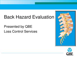
BACK EVALUATION Sara Brown, D.O. Assistant Professor Dept of Family Medicine CAQ Sports Medicine Loyola University Medical Center
RELEVANCE • Low back problems affect everyone at some time in their life • Yearly prevalence of 50% of working adults • 15-20% seek medical care • Fifth most common of PCP visits
ANATOMY Spine 5 Lumbar Vertebrae Sacrum Intervertebral discs Spinal cord Nerve roots
ANATOMY • Muscles • Latissimus Dorsi • Erector Spinae • Iliocostalis, Longissimus, Semispinalis • Quadratus Lumborum • Iliopsoas
“S’s” of the Spine Spondylolysis Pars interarticularis “Scottie dog” Spondylolisthesis Slippage of one vertebral body over another Scoliosis Curvature of spine Spinal Stenosis Narrowing of spinal canal
HISTORY Acute (<6 wks) MVA, Fall, Lifting,Twisting Subacute (6-12 weeks) Repetitive motion Posture Chronic (months/years) Workman’s comp, secondary gain Myofascial pain vs. organic etiologies Degenerative joint disease of the spine
HISTORY • Flexion-Based • Lumbar radiculopathy • Discogenic • Ruptured annulus fibrosis
HISTORY • Extension based • Spinal Stenosis • Spondylolysis • Spondylisthesis • Facet Syndrome
HISTORY • Either (Flexion and/or extension) • Muscular (myofascial) • Mechanical LBP • Sacroiliac (SI) joint • Osteoarthritis
AGE • Age <20 • Pars interarticularis stress fx • Spondylolisthesis • Scoliosis • Muscular • SI joint
AGE • Age 20-50 • Muscle strain (myofascial) • Mechanical LBP (inflexibility/imbalance) • Herniated disc • Sacroiliac • Facet syndrome
AGE • Age >50 • Herniated disc • Mechanical LBP (inflexibility/muscle imbalance) • Spinal Stenosis • Osteoarthritis • Facet arthropathy • Spondylisthesis (degenerative) • Compression fractures
RED FLAGS • Age >50 or <20 • History of cancer, weight loss and/or night pain - Tumor • Bowel/bladder probs or saddle anesthesia • Cauda equina • Weakness • Worsening radiculopathy • Fever/chills • Osteomyelitis • Pyelonephritis • Stiffness (Bamboo spine) • Inflammatory • Ankylosing spondylitis
PHYSICAL EXAM • Posture • Lumbar lordosis • Pelvic Height • Lumbopelvic rhythm • ROM • Flexion/Extension • Rotation • Sidebending • Severe guarding in all planes is a red flag
PALPATION • Spinous processes • Sacroiliac joints • Paraspinal muscles • Piriformis/Gluteus medius
NEUROLOGIC EXAM • Gait • Heel (L5) • Tip Toe (S1) • Strength • Hip flexion (L1) • Hip abduction (L2) • Quadriceps (L3) • Anterior tibialis (L4) • FHL/Abduction hip (L5) • Plantar flexion/Eversion (S1)
NEUROLOGIC EXAM • DTR’s • Knee (L4) • None for L5 • Ankle/Achilles (S1) • Sensation • L4 – medial foot • L5 – dorsal foot • S1 – lateral foot
PROVOCATIVE TESTING • One leg hyperextension • Spondylolysis • Straight leg raise or slump test • Supine/seated • Neural tension • Discogenic/radiculopathy • Pain below the knee at less than 70 degrees of flexion and aggravated by dorsiflexion most suggestive • Crossover pain is a stronger indication
PROVOCATIVE TESTING • FABER’s • Hip or SI Joint • Gainslen’s • SI Joint
WADDEL’S SIGNS • Superficial, nonanatomic tenderness • Inconsistent responses – positive straight leg raise, but negative slump test • Nondermatomal sensory loss • Over-reaction • No effort
IMAGING • In the absence of red flags, no imaging necessary initially • 90% resolve spontaneously in 4-6 weeks • Imaging studies on “normal” asymptomatic people are commonly abnormal
MANAGEMENT • Pain Control Tylenol NSAIDS Muscle relaxants Opiods Antiepileptics • Therapy based on diagnoses: • Flexion based pain • centralize pain with extension program (McKenzie) • Extension based pain • William’s flexion exercises
MANAGEMENT • Avoid bed rest • Heat/cold • Spinal manipulation • Massage therapy • Proper lifting mechanics - Hold close to body at level of navel - No twisting/bending/reaching while lifting • Ergonomics - Soft support for small of back, arm rests, etc • Acupuncture
EVIDENCE FOR ACUTE LBP NONPHARMACOLOGIC EFFICACY • Heat • Spinal manipulation
EVIDENCE FOR SUBACUTE LBP NONPHARMACOLOGIC EFFICACY • Intensive interdisciplinary rehabilitation • Exercise therapy • Acupuncture • Massage therapy • Spinal manipulation • Yoga • Cognitive-behavioral therapy • Progressive relaxation
COMMON DIAGNOSES • Discogenic • Flexion based pain • Leg pain>back pain if radicular PE Flexion pain + SLR, +/-neurologic Rx PT: McKenzie exercises Steroids/NSAIDs/antiepileptics Epidural steroids for leg pain Surgical decompression
MUSCULAR/MECHANICAL LBP • History • Stiffness in all planes • +/- h/o trauma • PE • Paraspinal muscle spasm • Inflexibility • Nl provocative testing • Rx • PT for core strengthening and teach proper posture & lifting mechanics • NSAIDs/muscle relaxants
SACROILIAC JOINT • History • Twisting/torque • +/- trauma • Deep, vague back or pelvic pain • PE • No pain above L5 • Nl ROM, neurologic • + FABER’s/Gainslen’s • Rx • NSAID’s • PT: pelvic stabilization and core strengthening • Manipulation • SI Joint injections
PARS STRESS FRACTURE • History Repetitive hyperextension Adolescents • PE + 1-leg hyperextension Nl neurologic, strength • Rx Limit extension activity Bracing PT (spinal stabilization)
SPINAL STENOSIS • History • Extension pain • Pain with walking, relieved by rest/flexion • PE • Flexed posture • +/- neurologic exam • Rx • Steroids/NSAIDs/antiepileptics • Flexion based therapy • Transforaminal/selective injections (flouroscopy)
FACET SYNDROME • History • Extension based pain • No leg pain • PE • Pain with extension • Nl neuro, strength • Nl provocative testing • Rx • NSAIDs • Flexion based therapy • Facet injections (flouroscopy)
MANIPULATION • Pelvic obliquity • SI joint pain • Mechanical low back pain • Muscular tension • Scoliosis • Postural pain
SOFT TISSUE • Palpate spinous processes • Place thenar and hypothenar eminences of dominant hand just lateral to spinous processes with other hand on top used as support • Press down first and then gently push laterally • Repeat this the length of the lumbar and thoracic spine on both sides
COUNTERSTRAIN • Find a tender point in the low back • Keep one finger on the point • Use your other hand to shorten the muscle by elevating the leg • Move the leg into different positions while monitoring the point to feel where it is the least tense • Hold for 1-2 minutes and monitor for release
MUSCLE ENERGY • Place thenar and hypothenar eminences of one hand just superior to the ilium on the side that you are standing on. • Use your other hand to extend the leg on that side to the natural barrier • Then have patient push down towards the table for 3 seconds • Relax for 1 second and extend the leg further to the new barrier • Repeat 3 times