Download
1 / 1
30 likes | 164 Vues
Pore Structure in Activated Carbon with Applications to Methane Storage Mikael Wood, Jacob Burress, Peter Pfeifer Department of Physics and Astronomy, University of Missouri-Columbia Parag Shah, Galen Suppes Department of Chemical Engineering, University of Missouri-Columbia. Introduction:

E N D
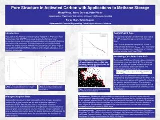
Pore Structure in Activated Carbon with Applications to Methane Storage Mikael Wood, Jacob Burress, Peter Pfeifer Department of Physics and Astronomy, University of Missouri-Columbia Parag Shah, Galen Suppes Department of Chemical Engineering, University of Missouri-Columbia Introduction: As a part of the Alliance for Collaborative Research in Alternative Fuel Technology (ALL-CRAFT) our group studies the formation and properties of pore structures in activated carbon with the goal of storing methane at low pressures. To study the pore structure of activated carbon we employ various methods including small/ultra-small angle x-ray scattering (SAXS/USAXS), methane and nitrogen adsorption, and electron microscopy. • SAXS/USAXS Data: • SAXS data indicate a predominate pore radius of ~20Å; in excellent agreement with nitrogen data. • SAXS data shows that sample BS-18 has a surface fractal dimension Dsurface= 2.3. This fractal behavior extends over a large range of r values and has an inner cutoff near 700Å. Scattering Calculated from PSD: To compare SAXS and nitrogen data we calculate a scattering curve from the PSD. To do this we assume an ideal two phase model in which the pores are spherical and uncorrelated. The scattering curve is then given by: Where V’(R) is a piecewise cubic Hermite interpolating polynomial fit through the PSD, V(R) is the volume of a sphere with radius R, and p0 is the electron density of carbon. The scattering curve produced from the PSD for sample BS-18 is compared to the experimental scattering of BS-18 in figure 3. Figure3: Experimental scattering (blue), calculated scattering from PSD (red), and calculated scattering with added power law (magenta) for sample BS-18. Figure 1: Pore size distribution from sample BS-18. The predominant pore size is ~19Å. Figure 2: Nitrogen adsorption (red) and desorption (blue) isotherms for sample BS-18. Figure 4: Cartoon of surface fractal penetrated by network of spherical pores. Nitrogen Sorption Data: Applying BJH analysis to data obtained from the nitrogen desorption isotherm for a given sample we are able to acquire a pore size distribution (PSD). We are also able to obtain other important information such as the total surface area per gram, micropore surface area per gram, and micropore volume. However, nitrogen data can not give us detailed information about the spatial distribution of the pores. To acquire spatial information about the pore structure we must turn to SAXS analysis. Conclusions: We see that the calculated and experimental curves in figure 3 agree quite well, especially at high q values (small R values). We, therefore, conclude that our sample is characterized by two separate regimes. Above 700Å it is a surface fractal with Dsurface= 2.3. At smaller R values we find uncorrelated spherical pores that scale in a non-fractal manner. A cartoon a such a situation is seen in figure 4. Such a detailed knowledge of the pore structure could not have been accurately acquired from nitrogen sorption or SAXS alone. Acknowledgments: This research is based on work supported by the National Science Foundation, under Grant No. EEC-0438469, the University of Missouri, the Department of Education (GAANN), and the Midwest Research Institute. Use of the Advanced Photon Source was supported by the U.S. Department of Energy, under Contract No. W-31-109-Eng-38.