Investigating Cytochalasin B Effects on Actin Cytoskeleton & Cell Viability in Mouse Fibroblasts
140 likes | 168 Vues
Explore the impact of Cytochalasin B on actin cytoskeleton in fibroblast cells using two assays—Actin staining and Cell viability. Each group will assay different CB concentrations for 1 hour. Utilize advanced techniques like flow cytometry and fluorescence microscopy to analyze results. Learn about cytoskeleton structure, molecular labeling, and flow cytometry protocols.

Investigating Cytochalasin B Effects on Actin Cytoskeleton & Cell Viability in Mouse Fibroblasts
E N D
Presentation Transcript
Cytoskeleton Lab 2:The Effects of Cytochalasin B on the Actin Cytoskeleton & Cell Viability in Mouse Fibroblast Cells
Today: Two Assays • Actin staining • Each group will assay two concentrations of cytochalasin B (CB) between 2 and 20 µM and will also do a control (no drug). • Cells treated for 1 h. • Cell viability (flow cytometry) • Each group will assay one of the two CB concentrations tested in the actin staining experiment (or a control with no drug). • Will assay cells treated 1 h or 24 h with the assigned concentration of drug (24 h samples started yesterday). • Data for the entire class will be pooled for your lab report Results.
Before the Lab Intro:• Obtain a 10-ml aliquot of culture medium containing 20 µM cytochalasin B (CB). Prepare 8 ml of medium containing your assigned CB concentration for the cell viability assay and 3 ml of medium containing your other assigned CB concentration for the actin staining assay.• Obtain one 4-chamber growth slide containing cells; dump medium from chambers into waste beaker (in hood). Label frosted portion of slide with your initials.• Add 1 ml fresh (drug-free) medium to 1st 2 wells of each slide (closest to frosted part of slide); add 1 ml of high CB medium to next well and 1 ml of low CB medium to last well.• Obtain one flask of cells diluted last week. Aspirate the culture medium and add 5 ml of medium containing your assigned CB concentration for the viability assay.• Incubate both slide and flask 1h in tissue culture incubator (37°C, 5% CO2).
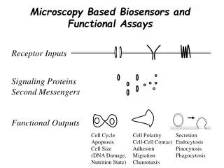
The Actin Cytoskeleton • Microfilaments formed from actin monomers • Filaments have polarity: plus & minus (or “pointed” & “barbed”) ends • Actin monomers add preferentially to plus/barbed ends
Cytochalasin B Effects on Actin: Effects on Cells:
Molecular labeling techniques used for fluorescence microscopy • Direct: fluorescent molecule binds directly to target molecule. • Ex: phalloidin conjugated to rhodamine (red fluorescent dye) binds specifically to F-actin, the filamentous form. • Indirect: fluorescent molecule binds to some other molecule used for recognition of the target molecule. • Untagged primary antibody used to recognize target protein & dye-conjugated secondary antibody used to visualize.
Amanita phalloides“Angel of Death” mushroom Phalloidin Phalloidin locks F-actin subunits of actin together, stabilizing actin filaments.
Actin stained with Phalloidin conjugated to FITC or Rhodamine
Flow Cytometry • Technique for measuring characteristics of cells (e.g. fluorescence, size, “granularity”) in a population on a cell-by-cell basis • Cells flow through a capillary in “single file”, passing through a laser beam of a certain wavelength. • The scattered light and fluorescence intensity are recorded for each cell. Fluorescence Activated Cell Sorting (FACS)—sophisticated flow cytometry
ViaCount Assay • Determines % of living and dead cells in a culture • All cells stain with a DNA dye (distinguishes from non-cell debris) & dead cells only stain w/ an exclusion dye (similar to Trypan Blue); two dyes emit fluorescence of different wavelengths • Flow cytometer quantifies intensity of fluorescence of at the two wavelengths as each cell passes through the laser Sample Data:
Protocol Steps after Drug Incubation Actin Staining Viability Assay PBS washes (4 x 3 min) Rinse cells w/ PBS Formaldehyde fixation (10 min) Trypsinize (30 sec + 15 min) Resuspend in 5 ml fresh medium PBS washes (3 x 3 min) Place 200 µl each cell suspension in a regular microfuge tube Cold acetone permeabilization (3 min) Wait for entire class to be ready PBS washes (3 x 3 min) Rhodamine-phalloidin staining (20-25 min) Mix cells with 2 µl 0.5X ViaCount reagent PBS washes (4 x 3 min) Do flow cytometry during rhodamine-phalloidin incubation Mounting medium, coverslip & seal
For Next Week (Week of April 16): • Sign up for microscope time slot • Bring research article for imaging method presentation to your microscope appt. for approval. For Week of April 30: • 10-15 min group presentation on chosen imaging topic & its application in your chosen research paper For Week of May 7: • Mini-lab report due (Title, Abstract, Results, References)