Download
1 / 122
1.24k likes | 1.52k Vues
Learn about skin and soft tissue infections, their clinical presentations, diagnosis, and management, including bacterial and viral causes. Understand the structure of human skin and normal skin microbiota. Explore various skin lesions and infections caused by pathogens such as Staphylococcus and Streptococcus. Discover treatment options and prognostic factors for severe infections.

E N D
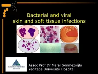
BACTERIAL and VIRAL SKIN AND SOFT TISSUE INFECTIONS ONUR OK HELİN YILMAZ
Skin and soft tissue infections (SSTIs) • SSTIs which include infections of skin, subcutaneous tissue, fascia, and muscle, encompass a wide spectrum of clinical presentations, ranging from simple cellulitis to rapidly progressive necrotizing fasciitis. Diagnosing the exact extent of the disease is critical for successful management of a patient of soft tissue infection.
The Structure of Human Skin Figure 21.1
Contact Dermatitis Necrotizing Fascitis Cellulitis Erysipelas Erythema Multiforme Ecthyma Deep Vein Thrombosis Folliculitis Impetigo
superficial Impetigo Ecthyma Folliculitis Furuncule/Carbuncle/Abscess Erysipelas Cellulitis Necrotizing Fascitis deep Stevens, DL, Bisno, AL, Chambers, HF, et al. Practice guidelines for the diagnosis and management of skin and soft tissue infections: 2014 update by the Infectious Diseases Society of America. Clin Infect Dis. 2014.
Normal Microbiota of the Skin • Gram-positive, salt-tolerant bacteria • Staphylococci • Micrococci • Diphtheroids Figure 14.1a
Normal Microbiota of the Skin • Grow on oils • Aerobes on surface • Corynebacterium xerosis • Anaerobes in hair follicles • Propionibacterium acnes • Yeast • Malassezia furfur
Skin Lesions Figure 21.2
Staphylococcal Skin Infections • Staphylococcus epidermidis • Gram-positive cocci, coagulase-negative • Staphylococcus aureus • Gram-positive cocci, coagulase-positive Clinical Focus, p. 593
Staphylococcus aureus • Antibiotic resistant • Leukocidin • Resists opsonization • Survives in phagolysosome • Lysozyme resistant • Exfoliative toxin • Superantigen Clinical Focus, p. 593
Staphylococcal Biofilms Pseudomonas,Enterobacter,Flavobacterim, Alcaligenes, Staphylococcus Figure 21.3
Staphylococcal Skin Infections • Folliculitis: Infections of the hair follicles • Sty: Folliculitis of an eyelash • Furuncle: Abscess; pus surrounded by inflamed tissue • Carbuncle: Inflammation of tissue under the skin • Impetigo: crusting (nonbullous) sores, spread by autoinoculation
Nonbullous Lesions of Impetigo Figure 21.4
Scalded Skin Syndrome • Toxic shock syndrome (TSS) • Toxic shock syndrome toxin 1 • Scalded skin syndrome • Bullous impetigo • Impetigo of the newborn
Lesions of Skin Syndrome Figure 21.5
Streptococcal Skin Infections • Streptococcus pyogenes • Group A beta-hemolytic streptococci • Hemolysins • Hyaluronidase • Streptolysins • M proteins
Group A Beta-Hemolytic Streptococci Figure 21.6
Ecthyma • Presentation: Vesicle/pustule which enlarges over several days and becomes thickly crusted. When crust is removed a superficial saucer shaped ulcer remains with elevated edges. • Nearly always on shins or dorsal feet. • Heals in a few weeks with scarring. • Agent: Staph or Strep. • Heal with scaring • Gangrene in predisposed individuals. • Treatment: Clean, topical and systemic ABX.
Scarlet Fever • Presentation: 24 –48 hrs after Strep. Pharyngitis onset. • Cutaneous: • Widespread erythema with 1-2 mm papules. Begins on neck and spreads to trunk then extremities. • Pastia’s lines – accentuation over skin folds with petechia. • Circumoral pallor • Desquamation of palms and soles at appox two wks. • May be only evidence of disease. • Other: strawberry tongue • Causes: erythrogenic exotoxin of group A Strep. • Culture to recover organism or use streptolysin O titer if testing is late. • TX: PCN, E-mycin, Cloxacillin.
Streptococcal Skin Infections • Necrotizing fasciitis • Erysipelas Figure 21.7
Necrotizing Fasciitis • Presentation: Following surgery or trauma (24 to 48 hours) - erythema, pain and edema which quickly progress to central patches of dusky blue discoloration. Anesthesia of the involved skin is very characteristic. By day 4-5 the involved area becomes gangrenous. • Infection of the fascia. • Many causative agents. Aerobic and anaerobic cultures should be taken. • Treatment: Early debridement. ABX. • 20% mortality in best cases • Poor prognostic factors: Age >50, DM, Atherosclerosis, involvement of trunk, delay of surgery >7 days.
Invasive Group A Streptococcal Infections • Exotoxin A, superantigen Figure 21.8
Erysipelas • Presentation: erythematous patch with a distinctive raised, indurated advancing border. Affected skin is very painful and is warm to touch. Freq. associated with fever , HA and leukocytosis >20,000. • Face and Legs are most common sites. • Involves superficial dermal lymphatics • Cause: Group A strep., (Group B in newborns) • Differential: • Contact derm: more itching little pain. • Scarlet fever: widespread punctate erythema • Malar rash of Lupus and Acute tuberculoid Leprosy: Absence of fever pain and leukocytosis. • Treatment: Systemic PCN for 10 days.
Streptococcal Toxic Shock Syndrome • M proteins • Complex with fibrinogen • Binds to neutrophils • Activates neutrophils • Release of damaging enzymes • Shock and organ damage
Infections by Pseudomonads • Pseudomonas aeruginosa • Gram-negative, aerobic rod • Pyocyanin produces a blue-green pus • Pseudomonas dermatitis • Otitis externa, or “swimmer’s ear” • Post-burn infections • Opportunistic • Hot-tub folliculitis
Infections by Pseudomonads • Hot Tub Folliculitis Hot tub folliculitis is an infection of the hair follicles caused by the bacteria Pseudomonas aeruginosa. This bacteria is commonly found in contaminated whirlpools, hot tubs, water slides, physiotherapy pools, or even loofah sponges
Folliculitis and Furuncle • FolliculitisFolliculitis is an infection that is localized to the hair follicle. A folliculitis looks like small, yellow pustules that are confined to the hair follicle. • FuruncleA furuncle is an infection of the pilosebaceous unit, therefore is more extensive than a folliculitis because the infection also involves the sebaceous gland. • Frequently occurs on the neck, face, armpits, and buttocks.