Download
1 / 40
400 likes | 535 Vues
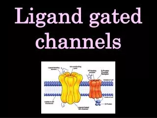
Second-Messenger Gated Ion Channels. Membrane Biophysics, 2014 Ion Channel Presentation Vehpi Yildirim and Joe McKenna. Overview. Stimulus triggers iIntracelluar signal that modulates channel activity Examples G-protein coupled channels IP 3 -regulated channels

E N D
Second-Messenger Gated Ion Channels Membrane Biophysics, 2014 Ion Channel Presentation Vehpi Yildirim and Joe McKenna
Overview Stimulus triggers iIntracelluar signal that modulates channel activity Examples G-protein coupled channels IP3-regulated channels Adenine nucleotide-sensitive channels
Examples G-protein coupled inward rectifying K+ channel IP3-regulated Ca2+ release from ER
ATP-sensitive K+ Channels Link cellular energetics and excitability Gate efflux of K+ Inward rectifier Shallow voltage-dependence Inhibited by ATP, activated by Mg2+
KATP Architecture Functional Octamer Kir 6.2: 4 sub-units Channel pore Site of ATP inhibition Sulphonylurea Receptor (SUR): 4 sub-units Site of Mg2+ activation
Kir 6.2/SUR Model Extrapolated from Bacteria K+ channel crystal Prokaryotic Kir Targeted mutation ATP binds at interface of SUR NBF1 & 2 (b. green)
Mechanism of Gating Fast ligand-independent gating by ion selectivity filter Ligand-dependent gating by hinged motion of M2 Inhibited by ATP Activated by PIP2, MgADP
Gating Kinetic Model Fast ligand-independent gating and slow ligand-dependent gating One subunit in closed configuration → channel closed Two ways to achieve same half-maximal inhibition
KATP-related disease Pancreatic beta-cells Loss of function mutation →Hyperglycemia/diabetes Gain of function mutation → Hyperinsulinemia Coronoary cells Loss of function mutation → spontaneous contraction, early death
Identification and Properties of an ATP-Sensitive K+ Current in Rabbit Sino-Atrial Node Pacemaker Cells X. Han, P. E. Light, W. R. Giles and R. J. French Journal of Physiology (1996), 490.2, pp.337-350
INTRODUCTION • K(ATP) channles have been identified in many cell types. • Most studies use myocytes from atrium. • Here they use cells from sino-atrium node.
Questions to be Answered • Are K(ATP) channels present in SA node and, if so, what are their single channel properties? • Can physiological, pharmacological and pathological conditions which are known to activate K(ATP) channels alter SA-node activity?
METHODS • Isolated single cells from SA node of rabbit heart are studied by measuring spontaneous activity. • Both whole cell and single channel currents are measured. • Pharmacological blockers or openers are used.
Ventricular myocytes also isolated to compare results from different regions of heart. • Perforated patch technique for Whole cell. • Inside-out configuration for single channel.
Glibenclamide: K(ATP) channel blocker. Acts on SUR subunit. Cromakalim and Pinacidil: K(ATP) channel openers. Act on SUR subunit.
Effects of glibenclamide on electrical activity and ion curents.
Effect of metabolic inhibition by NaCN NaCN (Sodium Cyanide) : inhibits ATP production.
Effects of drugs on single channel activity. Effects with high ATP concentration.
Neonatal Diabetes (NDM) Overview Presents within first 3 months of life, requires insulin treatment Insulin response to sulphonylureas but not glucose or glucagon May result from Kir 6.2 gain of function mutations in pancreatic beta-cells
KATP Channels and NDM Glucose → ATP → channel closure → Ca2+ influx → Insulin secretion
NDM Patient Screening Patients with known diabetes-related mutations excluded Physical exam including insulin, sulphonylurea challenges Kir 6.2 gene sequenced Identified 6 novel mutations NDM seen only in patients with Kir6.2 mutations
Kir6.2 Affected Residues Highly conserved regions → functional role Near ATP-binding site or slide helix
Patient Response to Secretagogues 3 patients with mutations in ATP binding site (ABS) No secretion from glucose Secretion from KATP channel opener
KATP Channels in Oocytes Channels with mutated ABS residues Larger current in steady [ATP] Current increased by sulphonylurea Weakly inhibition by ATP
KATP Channels in Oocytes NDM pathology more severe in homozygote mutants Significant difference in half-maximal activation by ATP
Conclusion Activating mutations in Kir6.2 causes NDM Found in 34% of patients with NDM Accompanying complications point to vital role of KATP channels in brain and muscle Potential therapy: channel blocker acting on SUR receptors
Defective Insulin Secretion and Enhanced Insulin Action in KATP Channel Deficient Mice Takashi Miki, Kazuaki Nagashima, FumiTashiro, Kazumi Kotake, Hideyuki Yoshitomi, Atsuko Tamamoto, TohruGonoi, Toshihiko Iwanaga, Jun-ichi Miyazaki, And Susumu Seino PNAS Vol. 95, pp. 10402-10406, September 1998, Biochemistry
INTRODUCTION • KATP Channels in pancreatic Beta Cells comprise Kir6.2 and SUR1 subunits. • KATP Channels are ATP and ADP sensors and play a very important role in insulin secretion. • Mutations in regulatory genes cause hypoglycemia. • Here they use Kir6.2-/- mice to study the role of KATP channels in insulin secretion.
Kir6.2+/+ and Kir6.2-/- cells are dialyzed with ATP-free pipette solution.
Glucose or Tolbutamide does not effect [Ca] in Kir6.2-\- cells. AcetylCholine and High K+ does effect [Ca] like in wild type cells. Showing voltage gated Ca channels and IP3 sensitive Ca stores are functioning normally in Kir6.2-\- cells.
A rapid rise in Ca concentration is needed for glucose induced insulin secretion, rather than a continuous elevated [Ca]. In Kir6.2-/- mice, only a small first phase and no second phase secretion observed. (In Vitro)
Glucose induced insulin secretion is reduced in knock-out mice. But surprisingly glucose lowering effect of insulin is significantly increased in knock-out mice.
Beta Alpha Kir6.2+/+ Kir6.2-/-
CONCLUSION • KATP channels play a significant role in insulin secretion. • Glucose metabolism itself is insufficient for glucose-induced and sulfonylurea-induced insulin secretion, both of which require the rapid rise in [Ca2] caused by closure of the KATP channels.