
Portal Venous Aneurysm in Chronic Pancreatitis: A Rare Encounter
Explore the unique association between splenic vein aneurysm and chronic pancreatitis, including diagnosis, treatment, and follow-up recommendations. Learn from a clinical case and relevant literature reviews.

Portal Venous Aneurysm in Chronic Pancreatitis: A Rare Encounter
E N D
Presentation Transcript
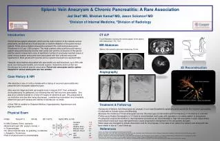
Splenic Vein Aneurysm & Chronic Pancreatitis: A Rare Association Jad Skaf1 MD, Bhishak Kamat2 MD, Jason Solomon2 MD 1Division of Internal Medicine, 2Division of Radiology • Introduction • Portal venous system aneurysm, which are the most common of the visceral venous aneurysms, are defined as a focal saccular or fusiform dilatation of the portal venous system. Portal venous system aneurysms represent 3% of all venous aneurysms. Prevalence is 0.6 per 1,000 persons. The most common sites at which portal venous system aneurysms develop are the main portal vein and the confluence of the splenic and the superior mesenteric veins. A significant number of previously reported cases of portal venous system aneurysms were associated with liver cirrhosis and portal hypertension. Most people with a portal venous system aneurysm are asymptomatic. • Vascular abnormalities associated with pancreatitis are well described, up to 50% with acute necrotizing pancreatitis, and include superior mesenteric and/or portal vein thromboses and arterial pseudo-aneurysms. Portal vein aneurysms and/or spleno-mesenteric venous aneurysms are rare entities. CT A/P Focal Dilatation involving the central aspect of the splenic vein measuring 1.7x1.7 cm. MR Abdomen Splenic Vein pseudo-aneurysm measuring 18 mm. 3D Reconstruction Angiography Case History & HPI • We describe a case of a 68 yo female with a history of recurrent pancreatitis who presented with intractable abdominal pain. • She was first diagnosed with pancreatitis back in August 2007, then underwent cholecystectomy for gallstones, but following that she had recurrent pancreatitis. She was at an outside hospital for a total of 5 weeks for abdominal pain. She presented to our institution one day after being discharged , complaining of persistent and intractable abdominal pain with nausea and inability to tolerate per os intake. • Other PMH is notable for Diabetes Mellitus, Hyperlipidemia, Hypertension and Hypothyroidism. Treatment & Follow-up • Symptoms of Splenic Vein Aneurysms are unusual. In our case the patient’s symptoms were caused by the recurrent pancreatitis. • Complications include rupture and Bleeding. • Because the incidence of these aneurysms are low, the exact type of intervention and the frequency of monitoring is unknown. • Follow-up by Duplex Sonography or CT-Scan is recommended every year until regression or a stable pattern is recognized. • Prophylactic surgical intervention or decompressive procedures are recommended in high-risk aneurysms in such cases where mechanical forces such as portal hypertension cause progressive enlargement of the aneurysms and pain. • Most cases are managed by simple observation and do not progress. In the case of our patient her splenic vein aneurysm has been stable in size at 1 year follow-up. Physical Exam • Vitals: Temp 97.2, HR 68, BP 149/72, SpO2 98% on RA • In Mild Distress (Pain), cachectic • Abdominal Exam: BS present normal, n • o Organomegaly. • No rebound tenderness, no guarding, no defense. • + Epigastric Tenderness. • Rest of physical exam is unremarkable Amylase 165 Lipase 134 Alkaline Phos. 77 Bilirubin Total 0.3 Bilirubin Direct 0.1 135 100 4 References 165 3.6 0.7 26 -Splenic vein Aneurysm with Calcification of Splenic and Portal Veins Ercan Kocakoc, MD *, Adem Kiris, MD, Zulkif Bozgeyik, MD, Hadi Uysal, MD, Hakan Artas, MD. Firat University Faculty of Medicine, Department of Radiology, Yahya Kemal Street, 23119, Elazig, Turkey -Splenic vein aneurysm—report of a lesion that progressively expanded Mo Ohhira, MD M Ono, MD Ma Ohhira, MD A Matsumoto, MD H Ohta, MD and M Namiki, MD. Department of Internal Medicine III, Asahikawa Medical College, Nishikagura 4–5–3, Asahikawa 078, Japan -Splanchnic vein aneurysms: a report of 13 cases. Weber G, Milot L, Kamaoui I, Pilleul F.J Radiol. 2008 Mar;89(3 Pt 1):311-6. Review. French. -Vascular disease of the spleen. Vanhoenacker FM, Op de Beeck B, De Schepper AM, Salgado R, Snoeckx A, Parizel PM. Semin Ultrasound CT MR. 2007 Feb;28(1):35-51. Review. 8.2 ALT 13 AST 22 Albumin 2.8 5.4 244 26.6


