Download

1 / 33
350 likes | 629 Vues
Acute Kidney Injury (AKI). Dr Svitlana Zhelezna Clinical Teaching Fellow UHCW NHS Trust svitlana.zhelezna@uhcw.nhs.uk 2013/2014 academic year. Objectives:. Recognise AKI Investigate and decide on: pre-renal, renal and post renal causes Recognise and manage hypovolemia

E N D
Acute Kidney Injury (AKI) Dr Svitlana Zhelezna Clinical Teaching Fellow UHCW NHS Trust svitlana.zhelezna@uhcw.nhs.uk 2013/2014 academic year
Objectives: • Recognise AKI • Investigate and decide on: pre-renal, renal and post renal causes • Recognise and manage hypovolemia • Manage hyperkalemia • Indications for emergency dialysis and heamofiltration
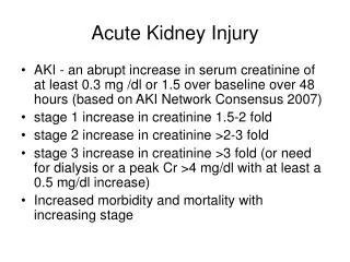
Definition of AKI • Rise in serum creatinine >50% from baseline • Or • Urine output <0.5ml/kg/hr for 6 hours
Effects of Acute Kidney Injury: Raised Urea, Creatinine and Uric Acid: - Confusion - Drowsiness Failure to Excrete Normal Acidic Products: - Metabolic Acidosis - Respiratory Hyperventilation Electrolyte Imbalances (Hyperkalaemia): - Dysrhythmias
Urea (2.5-7.5 mmols/L) Other causes for raised urea: • High protein intake. • Increased tissue breakdown (i.e febrile illness, crush injuries). • Dehydration. • Steroid or Tetracycline Administration. • Absorption of Blood fromG.I. Tract.
Creatinine (60 ‑125 mmols/L) • is a by product of normal muscle metabolism • is excreted in urine primarily as a result of glomerular filtration • more reliable indicator of renal function and of a glomerular filtration than Urea.
Acute Tubular Necrosis Renal tubular cell injury after a toxic or ischaemic insult results in: • sloughing of tubular debris and cells into the tubular lumen with eventual obstruction of the tubular flow, • increased intra‑tubular pressure and back leak of glomerular filtate out of the tubule and into the interstitium and renal venous blood
Three Phases of AKI: • Phase 1: Oliguric Phase. Usually lasts 10‑14 days but may last from several hours to several weeks. • Phase 2: Poliuric Phase. Occurs 2 to 6 weeks after the onset. • Phase 3: Recovery Phase. May last 3 to12 months.
AKI Outcomes: • Renal function loss – i.e. persistent loss of renal function lasting > 4 weeks • End Stage Kidney Disease – i.e. GFR < 15ml/min for > 3 months • Other associated complications – e.g. sepsis, bleeding, respiratory failure etc. • Increased Mortality
What to look for when clerking ? Ask about: • family history of renal disease • exactly when the presenting symptoms started, and which came first • joint pains, or rash, or nose bleed, or ear trouble (vasculitis) • backache or bone pains (myeloma and other malignancy) • drugs taken (NSAID, ACEI ect.)
Volume Status/Dehydration: • Skin Turgor • Mucus Membranes • JVP • Pulse rate and volume • Blood Pressure (check postural BP) • Peripheral perfusion –capillary refill • Chest sounds • Peripheral Oedema • Urine output
Investigations: • U&E’s, FBC, LFTs, ABG • Urine Dip/MSU if indicated • ECG • CXR • CRP if indicated • Blood cultures if indicated
Principles of Treatment: • Check Medication! Stop all nephrotoxic if you can (ACEI, diuretics, NSAIDS), Check that the dosages of those remaining /commencing are correct in renal failure (Enoxaparin, some antibiotics) • Treat lifethreatening hyperkalaemia first • Correct hypovolaemia/hypoperfusion – restore pressure • Exclude obstruction ASAP (Imaging) • Treat the underlying cause • Consider Renal replacement therapy if no response
Case 1 • 66 y.o. man presents to A&E at 10am • PC: increasing SOB for 7/7, coughing up phlegm and having fever. • PMH: DM, HTN • DH: metformin, aspirin, ramipril, atenolol and simvastatin. • O/E: pale, sweaty, BP 85/50, HR 115, Sats 92% on air, RR 25, T 38.3, coarse crackles on the right side of his chest. • CXR - RLL pneumonia. • Blood results: Na 130, K 4.5, Urea 14.3, Creat 189 The nurse asks you to assess the patient at 2 pm as he hasn't passed urine since admission. What would be your management?
Initiate management: • Reassess the patient including volume status, vitals, check the catheter if in place • CHECK CURRENT MEDICATIONS! • Investigations: ABG, Urine dip • Treatment: fluid resuscitation, call for senior help
Fluid balance (adults, resting state, mL per day) Totaling: in/out ~2500 ml/day
Maintenance fluids: WEIGHT RATE For the first 10 Kg 100 mL/kg/24hrsor 4 mL/kg/hr For the next 10-20 Kg Add 50 mL/kg/24hrsor +2 mL/kg/hr For each Kg above 20 Add 20 mL/kg/24hrsor +1 mL/kg/hr So, the maintenance fluid requirements for a 25-kgchild is: 1000 + 500 + 100 = 1600 (mL/24hrs) or 40 + 20 + 5 = 65 (mL/hr) So, the maintenance fluid requirements for a 70-kgadult is 1000 + 500 + 1200 = 2700 (mL/24hrs) Or 40 + 20 + 50 = 110 (mL/hr)
Pre-existing Normal Deficits: (missing maintenance) is estimated bymultiplying the normal maintenance volume by the length of the fasting period: For 70‑kg man fasting for 8 hours this amount is (40 + 20 + 50) mL/h x 8 hrs = 880 mL. In reality, this deficit will be somewhat less as a result of renal conservation EstimatedAbnormal Fluid Losses: Known losses: bleeding, vomiting, excessive diuresis or diarrhoea… Occult losses due to fluid sequestration in body cavities or traumatized tissues (obstructed bowels, ascites, intramuscular haematoma …); Increased insensible losses due to hyperventilation, fever and sweating (an extra 500 ml/day is required for every degree Celcius above 37, ~20 ml/hr); Fluid requirements in illness:
Fluid requirements in illness Maintenance requirements for an adult Na - 50-100 mmol/day K - 40-80 mmol/day In 1.5-2.5 Iitres of water by the oral, enteral or parenteral route (or a combination of routes). Additional amounts should only be given to correct deficit or continuing losses
Contents of common crystalloids in mmol/L Na K Ca Cl HCO3 Osm pH Plasma 140 4 2.3 100 26 285-295 7.4 Na Cl 0.9% 154 0 0 154 0 308 5.0 Dextrose 5% 0 0 0 0 0 252 4.0 Dex.Saline 30 0 0 0 0 255 4.0 Hartmann’s 131 5 2 111 0 278 6.5 Lactate 29 Ringer’s 147 4 2.2 156 0 302 6.9 Lactate 28 Na Bicarb 1.2% 150 0 0 0 150 300 8.0 Na Bicarb 8.4% 1000 0 0 0 1000 2000 8.0
Fluid requirements in illness Excessive losses from gastric aspiration/vomiting crystalloid solution with K supplement. ↓Cl - 0.9% NaCl + K (sufficient amount) and care not to produce sodium overload. ↓Na (excessive diuretic exposure) -Hartmann's Diarrhoea, ileostomy, small bowel fistula, ileus, obstruction - volume for volume with Hartmann's .
What is Hyperkalaemia? Level of potassium above 5.5 mmol/l in venous blood ECG changes (peaked T waves and broadening of QRS complex) are important but may NOT be seen even if potassium level is life threatening May cause sudden death or progressive bradycardia and death
Causes of Hyperkalaemia: • AKI/Renal failure • Sepsis with acute kidney injury • Drugs (spironolactone, ACE inhibitors, amiloride and OTHERS)
Acute Renal Failure →Emergency Haemodialysis: • K+ > 7mmol/L, resistant to medical therapy • Pulmonary oedema refractory to medical therapy • Metabolic pH < 7.2 or base excess < -10 • Other possible indications include: • Uraemic pericarditis • Uraemic encephalopathy
Dialysis: No clear proven advantage for either in treatment of renal failure Theoretical advantage of clearance of middle molecules Haemofiltration: No need to transfer patient to renal unit Can be continuous Improved haemodynamic stability Permits vasopressers and other drug therapies including TPN Reduced risk of disequilibrium syndrome Renal Replacement Therapy
When to call nephrology? • Any known dialysis patient admitted • Any known renal transplant patient admitted • Any case of AKI where cause is not clear • Worsening AKI • Emergency dialysis indications • Suspect glomerulonephritis
Summary: • worry if • Patient has not passed urine or very little • U&E creatinine is going up, check dynamics • Patient is dehydrated plus cardiovascular compromised (past MI, CCF) • remember • Normal creatinine does not mean patient is not developing AKI • Call early for senior or specialist help
Thank you! Any questions?