Download
1 / 22
580 likes | 1.46k Vues
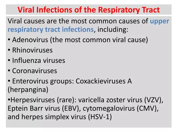
Viral Infections of the Respiratory Tract. Viral causes are the most common causes of upper respiratory tract infections , including: Adenovirus (the most common viral cause) Rhinoviruses Influenza viruses Coronaviruses Enterovirus groups: Coxackieviruses A (herpangina)

E N D
Viral Infections of the Respiratory Tract Viral causes are the most common causes of upper respiratory tract infections, including: Adenovirus (the most common viral cause) Rhinoviruses Influenza viruses Coronaviruses Enterovirus groups: Coxackieviruses A (herpangina) Herpesviruses (rare): varicella zoster virus (VZV), Eptein Barr virus (EBV), cytomegalovirus (CMV), and herpes simplex virus (HSV-1)
Adenoviruses: Name originates from Greek word “adenas” which means gland, site from which were initially isolated. Virions are icosahedral non-enveloped, 70-90 nm in diameter with double stranded DNA genome. Fibers protruding from capsid facilitate binding on to host 11 proteins are found in the virion Genome encodes for up to 50 proteins Divided into 6 groups (A to F) comprising 51 human serotypes
ni Incubation period: 5 – 8 days Transmission: Respiratory droplets, faecal- oral route, and contact either by hand to eye or sexual. Age affected: commonly among school-aged children They cause latent infection in the tonsils, adenoids and other lymphoid tissue. Diagnosis: Isolation of the virus from the respiratory or eye discharge or tissue culture. A fourfold or greater rise of antibody titre is a good evidence of infection.
a Clinical Picture: Respiratory diseases: pharyngitis, rhinitis, laryngitis or pneumonia Acute respiratory disease (ARD) among military recruits. Eye infection: conjunctivitis or keratoconjunctivitis Pharyngo- conjunctival fever Infantile gastroenteritis Acute haemorrhagic cystitis in children
Rhinovirus: Name originates from a Greek word means "nose“ They cause common cold or acute bronchitis Picornoviridae family Virions are icosahedral non-enveloped,single stranded RNA virus Over 100 serotypes are known Optimum growth occurs between 33 and 34 deg Celsius Not stable below the pH of 5-6
a Incubation period: 1 to 3 days (short) Transmission: droplet infection or hand-to-hand contact Multiplication of the virus: locally with no blood invasion Affected age: All ages The disease occurs sporadically or in epidemics. Secondary bacterial infection may cause otitis media, sinusitis, bronchitis or bronchpneumonia especially in children. Immunity is mainly superficial by IgA and interferon (short term immunity).
a Clinical picture: Human rhinoviruses are the primary cause of common colds. Symptoms include: sore throat, runny nose, nasal congestion, sneezing and cough; sometimes accompanied by muscle aches,fatigue, malaise, headache, muscle weakness, or loss of appetite.
Influenza Virus: Orthomyxoviridae family They replicate in mucous membrane of upper and lower respiratory tract They are helical, enveloped, single stranded RNA genome They are enclosed in a lipid envelop and a layer of glycoprotein spikes known as haemagglutinin (HA) and neuraminidase (NA) which are major antigenic determinants. They are divided into: types A, B, C on the basis of the nucleoprotein antigen Influenza occurs in epidemics and pandemics
HA - hemagglutinin NA - neuraminidase helical nucleocapsid (RNA plus NP protein) lipid bilayer membrane polymerase complex M1 protein ORTHOMYXOVIRUSES type A, B, C : NP, M1 protein sub-types: HA or NA protein
Role of H AND N Proteins H = Hemagglutinin and N = Neuraminidase Hemagglutinin allows the virus to bind to host cells Neuraminidase helps the virus to release itself from the highjacked cells in which it has reproduced Influenza A Virus Constantly Changes Antigenic drift Small changes in H or N proteins that occur from year to year Population is partially immune, but may be re-infected over time (periodic epidemics) Antigenic shift Acquisition of new H or N protein, possibly from an animal virus Population is not immune, everyone is susceptible (pandemics)
Influenza A Virus: Influenza A virus only is further classified into subtypes based upon HA and NA antigens 16 HA subtypes and 9 NA subtypes are now recognized circulating in birds, humans, swine and horses. The most famous subtypes are: A (H1N1): circulating in humans causing swine flu A (H5N1): circulating in birds causing avian flu Influenza B Virus: Infects mammals only Usually less severe illness
Clinical picture: Fever Headache Myalgia Cough Rhinitis Ocular Symptoms Incubation period: 1 to 2 days Transmission: droplet infection or hand-to-hand contact Diagnosis: 1- Isolation of the virus from nose, throat swab 2- Tissue culture 3- Provisional - clinical picture + outbreak
Coronaviruses: Name originates from a Greek word meaning crown because of the crown like appearance of the surface projections. Family Coronaviridae They are large helical, enveloped, single stranded RNA viruses The human coronaviruses (CoVs) are responsible for about 30% of mild upper respiratory tract illness (common cold) Newly emerged SARS-CoV causes severe acute respiratory syndrome (SARS) that has been reported in Asia, North America, and Europe.
SARS Disease: Incubation period: 2 - 10 days Transmission: droplet infection or contact to contaminated skin or fomites Clinical Picture: Fever, chills, rigors, headache, myalgia and malaise Respiratory symptoms often begin 3-7 days after symptom onset and peak in the second week. Laboratory Diagnosis: Serological Testing IFA: Indirect fluorescent antibody ELISA: Enzyme-linked immunosorbent assays Only for specimens obtained > 21 days by fever Molecular Testing RT-PCR: Reverse transcriptase-PCR Can detect infection within the first 10 days Culture:SARS-CoV (Vero E6 cell)
Herpesviruses: Icosahedral, envelopeddouble stranded DNA viruses. Genome consisits of long and short fragments which may be orientated in either direction, giving a total of 4 isomers. Three subfamilies: Alphaherpesviruses - HSV-1, HSV-2, VZV Betaherpesviruses - CMV, HHV-6, HHV-7 Gammaherpesviruses - EBV, HHV-8 Set up latent or persistent infection following primary infection Reactivation are more likely to take place during periods of immunosuppression Both primary infection and reactivation are likely to be more serious in immunocompromised patients.
Herpesvirus Particle HSV-2 virus particle. Note that all herpesviruses have identical morphology and cannot be distinguished from each other under electron microscopy
Herpes Simplex Viruse Type 1 (HSV-1): Double stranded DNA enveloped virus with a genome of around 150 kb The genome of HSV-1 and HSV-2 share 50 - 70% homology. They also share several cross-reactive epitopes with each other. There is also antigenic cross-reaction with VZV. Man is the only natural host for HSV. Transmission: By direct contact Primary infections usually involves the mucous membrane of the mouth Latency of HSV-1 is in trigeminal ganglia Recurrent lesions manifest at any site innervated by the affected neurons.
HSV-1 Disease: Acute Gingostomatitis: the commonest classic presentation Recurrent herpes labialis (cold sores) Encephalitis Keratoconjunctivitis Disseminated infections: e.g. pneumonia Diagnosis: 1- Isolation of virus on tissue culture 2- Detection of HSV in vesicle fluid by electron microscopy 3- Detection of viral DNA by PCR 4- Detection of viral antigen by direct immunofluroescence or ELIZA 5- Serological diagnosis to detect IgM antibodies that indicates recent infection or reactivation
HSV-1 Disease: Cytopathic Effect of HSV in cell culture: Note the ballooning of cells. Positive immunofluorescence test for HSV antigen in epithelial cell.
Varicella- Zoster Virus (VZV): This virus causes 2 diseases: (1) Chicken pox (Varicella): Infectious disease of children Characterized by fever and vesicular rash Transmission: respiratory route Vesicles appear first on trunk And then spread to face and extremities Recovery is the rule May be complicated with encephalitis
Varicella- Zoster Virus (VZV): (2) Herpes Zoster (Shingles): Infectious disease of adults Characterized by painful vesicular eruptions in areas of skin supplied by sensory nerves mainly thoracic & lumbar or trigeminal nerve. Results from reactivation of latent varicella infection in the neurons. May be complicate lymphoma, leukemia or immunosuppresion
Diagnosis of VZV: Virus Isolation: rarely carried out as it requires 2-3 weeks for a results. Direct detection: electron microscopy may be used for vesicle fluids. Immunofluorescense on skin scrappings can distinguish between the two. Serology: the presence of VZV IgG is indicative of past infection and immunity. The presence of IgM is indicative of recent primary infection. Cytopathic Effect of VZV in cell culture: Note the ballooning of cells.