Download
1 / 39
390 likes | 468 Vues
Explore the complex network of arteries, veins, and cerebrospinal fluid in the brain. Learn about the Circle of Willis, clinical considerations like stroke and hemorrhage, and the crucial role of the blood-brain barrier. Discover how cerebrospinal fluid provides mechanical protection for the brain and spinal cord.

E N D
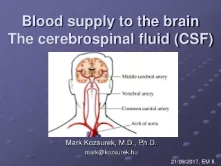
Blood supply to the brainThe cerebrospinal fluid (CSF) Mark Kozsurek, M.D., Ph.D. mark@kozsurek.hu 21/09/2017, EM II.
Extremlyhighdemandforoxygen and nutrients: human brain represents 2% of the body weight, but receives 15% of the cardiac output, 20% of total body oxygen consumption and 25% of total body glucose utilization. • Cerebrovasculardeseases and strokeareamongthe major causes of death.
2 sources of blood: ICA and VA
Vertebro-basilar system CTA: CT angiography atlas axis laterally upward backward C6
C7 cavernous sinus C6 C5 carotid canal C4 C3 ant. clinoid proc. C2 C1 foramen lacerum X-ray angiogram
C6 C5 C7 C4 (C3) cavernous sinus C2
Anotherclassification: Majority of these branches will never be seen and is not necessary to note them!
oculomotor n. abducens n. Circle of Willis pituitary stalk optic chiasm mamillary bodies
Circle of Willis encloses the optic chiasm, pituitary stalk and mamillary bodies. 2. Oculomotor nerve exits between the post. cerebral and sup. cerebellar arteries. 3. Vertebral arteries of the two sides unite to form the basilar artery at the ponto-medullary junction. The root of the abducens nerve and initial segment of the ant. inf. cerebellar artery can also be found here.
parietooccipital sulcus callosomarginal br. pericallosal br. A3 A2 A1 ant. communicating
parietooccipital sulcus ACA PCA MCA
oculomotor n. PCA sca BA aica VA pica sca: superior cerebellar aica: anterior inferior cerebellar pica: posterior inferior cerebellar
Clinicalconsiderations Atherosclerosis – braininfarctions Subarachnoidalhemorrhage
Ant. cerebral artery Weakness/paralysis of muscles and loss of sensory functions on the lower limbs of the contralateral side. Middle cerebral artery Paralysis and sensory disfunction involving head and neck and the upper limbs of the contralateral side. In case of damage of the dominant hemisphere speech disorders are also present. Post. cerebral artery Visual field defficiencies or blindness. Vertebro-basillar system Eye movement (gaze) disorders, double vision Anisocoria (pupils are different in size) Vertigo, loss of balance Dysphagia and dysphonia (disorder of swallowing and phonation) Drowsiness or unconsciousness
Blood-brainbarrier (BBB) • The extracellular fluid of the CNS is separatedfromthebloodbythe BBB ensuringstrictlycontrolled and mainlycarrier protein assistedtransport of macromolecules. • Is formedbyendothelialcellsattachedtooneotherbytightjunctions, basementmembrane, astrocyticendfeet. • Protectsthe CNS frompossiblytoxicagentsbutmakesdevelopment of medicinesactingonthe CNS difficult (e.g. antibioticsininfections).
Life outside the BBB: the circumventricular organs • „Circumventricular” = around the ventricles • Incomplet or missing BBB • Highly capillarized structure • Secretion of neurohormons or detection of hormons, glucose, ions, etc.
superior sagittal sinus SUPERFICIAL VEINS TROLARD’S VEIN LABBE’S VEIN cavernous sinus transverse sinus
DEEP VEINS ant. cerebral deep middle cer. v. of septum pell. choroid thalamostriate internal cerebral basal great cerebral vein
Almost thetotalvolume of veinousbloodcollectedfromthebrainleavestheskullthroughthejugularforamen and theinternaljugularvein. • Ifthejugularforamen and/ortheinternaljugularvein is gettingoccluded, bloodmayescapethroughthediploic and emissaryveinsconnectingtheduralsinuseswiththeveins of thescalpskin.
Diploic veins (frontal, anterior and posterior temporal, occipital): form a network between the external and internal compact bony layers of the skull and connect dural sinuses with the external veins.
emissary diploic Emissary veins (occipital, parietal, condylar, mastoid): pearce the skull directly and connect dural sinuses with external veins.
III. The cerebrospinal fluid (CSF) • Providesmechanicalprotectionforthebrain and thespinalcord. • Whenfloatinginthe CSF brainweightsonly 50g (!) accordingtotheArchimedes’ principle.
internal and external CSF spaces internal = ventricles external = subarachnoidal space
Choroid plexus of fourth ventricle Surface of a choroid plexus
post. choroidal from PCA ant. choroidal from ICA or MCA choroidal a. of the 4th ventricle from pica
1 median aperture of Magendi 2 lateral apertures of Luschka cerebellomedullary (or great) cystern lateral pontine (or pontocerebellar) cystern
Site of CSF resorption: arachnoid granulations in the superior sagittal sinus and lateral lacunae.
Hydrocephalus • Increasedvolume and/orpressure of CSF duetoacceleratedsynthesis, blockedcirculationorinsufficientresorption of liquor.
anterior cerebral middle cerebral posterior cerebral

![CEREBRAL CIRCULATION AND CEREBROSPINAL FLUID [CSF]](https://cdn2.slideserve.com/4005143/slide1-dt.jpg)