Download
1 / 346
3.65k likes | 4.53k Vues
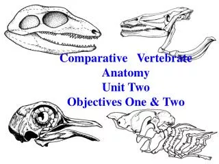
Comparative Vertebrate Anatomy Unit Two Objectives One & Two. Vertebrate Skeletal Functions. Gives the body shape. Supports the body weight. Acts as levers for movement. Provides surface for muscle attachment (origins & insertions). Vertebrate Skeletal Functions.

E N D
Comparative Vertebrate Anatomy Unit Two Objectives One & Two
Vertebrate Skeletal Functions Gives the body shape Supports the body weight Acts as levers for movement Provides surface for muscle attachment (origins & insertions)
Vertebrate Skeletal Functions Protects soft tissues and internal organs Mineral storage Blood cell production
Vertebrate Skeletal Components Exoskeleton - superficial or integumental * arises from the dermis or keratinized epidermis * provides protection for body surface & external support * dermal bone may sink and associate with deep bone to form a composite bone
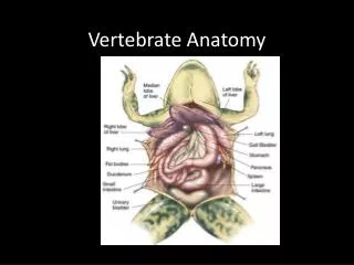
Vertebrate Skeletal Components Endoskeleton – deep bone * arises from the mesoderm * includes bones and associated cartilages & ligaments * supports and protects the body, provides surface for muscle attachment, produces blood cells and stores minerals
Vertebrate Skeletal Components The vertebrate skeleton can be divided into two regions: * cranial skeleton (skull) > splanchnocranium > chondrocranium > dermatocranium * functions to support and protect the brain and sense organs
Vertebrate Skeletal Components The vertebrate skeleton can be divided into two regions: * postcranial skeleton > axial skeleton > forms the longitudinal axis > appendicular skeleton > pectoral & pelvic girdles
Vertebrate Skeletal Components Splanchnocranium * arises from the ectodermal neural crest cells * forms the branchial arches * comprised of up to five articulated elements/side > pharyngobranchial, epibranchial, ceratobranchial, hypobranchial, basibranchial
Vertebrate Skeletal Components Splanchnocranium functions: * supports gills * surface for attachment of respiratory muscles * contributes to jaw (arch I) * contributes to hyoid arch (arch II)
Vertebrate Skeletal Components Chondrocranium * arises from the ectodermal neural crest cells and the mesenchymal cells of the mesoderm * the ectodermal elements form the nasal capsule and part of the otic capsule and trabeculae * the mesodermal elements form the rest of the chondrocranium
Vertebrate Skeletal Components Chondrocranium functions: * forms the braincase of elasmobranchs (no ossification) * forms the scaffolding of the brain in other vertebrates (partial to total ossification)
Vertebrate Skeletal Components Dermatocranium * arises from ectodermal and mesodermal tissues of the dermis * made up of dermal bones that are produced by intramembranous ossification * comprises most of the skull in amniotes
Vertebrate Skeletal Components Dermatocranium functions: * forms the roof and lateral elements of the skull, completing a bony encasement for the brain * contributes to the jaw casement
Vertebrate Skeletal Components Dermatocranium can by groups in “series” according to their location Facial series are the bones that encircle the external nares and form the snout > premaxilla > maxilla > nasal > septomaxilla (may be absent)
Vertebrate Skeletal Components Dermatocranium can by groups in “series” according to their location Orbital series are the bones that encircle the eye > lacrimal > prefrontal & postfrontal > postorbital > jugal
Vertebrate Skeletal Components Dermatocranium can by groups in “series” according to their location Temporal series are the bones that lie behind the orbit & complete the posterior wall of the braincase > intertemporal & supratemporal (absent in amniotes) > tabular (absent in amniotes) > squamosal > quadratojugal
Vertebrate Skeletal Components Dermatocranium can by groups in “series” according to their location Vault series are the bones that form a roof over the brain > frontal > postparietal (interparietal) > parietal
Vertebrate Skeletal Components Dermatocranium can by groups in “series” according to their location Palatal series are the bones that form the roof of the mouth (may bear teeth) > pterygoid > vomer > palatine > ectopterygoid > parasphenoid
Vertebrate Skeletal Components Dermatocranium can by groups in “series” according to their location Mandibular series are the bones that form the jaw > dentary > splenials > angular > surangular > prearticular > coronoids
Comparative Vertebrate Anatomy Unit Two Objective Three
Vertebrate Jaw Formation The jaw first formed in acanthodians and placoderms These fish were no longer confined to ciliary/mucus feeding Opened up an enormous variety of new food sources, first as a capture trap and then as a crushing, chewing structure
Vertebrate Jaw Formation The serial theory of jaw origin ~ T. H. Huxley ~ jaws developed from the first two pair of gill arches ~ arch I gave rise to the mandibular arch, while arch II gave rise to the hyoid arch ~ posterior arches became gnathostome branchial arches
Vertebrate Jaw Formation The composite theory of jaw origin ~ Erik Jarvik ~ hypothesizes ten original branchial arches ~ arch I » terminal arch ~ arch II » premandibular arch ~ arch III » mandibular arch ~ arch II » hyoid arch
Vertebrate Jaw Formation The composite theory of jaw origin ~ states that several elements of several arches came together to form one composite mandible
Vertebrate Jaw Attachment Jaw attachment types are based on their suspensorium (how the mandible is attached) Currently six recognized types of jaw attachment
Vertebrate Jaw Attachment Euautostylic - mandibular arch suspended by itself Paleostylic - no arches attached to the skull
Vertebrate Jaw Attachment Amphistylic - jaw attached by two articulations > anteriorly by a ligament to the palatoquadrate > posteriorly to the hyomandibula
Vertebrate Jaw Attachment Amphistylic
Vertebrate Jaw Attachment Metautostylic – jaw attaches directly through the quadrate bone Hyostylic – mandibular arch attaches through the hyomandibula
Vertebrate Jaw Attachment Craniostylic - lower jaw (dentary) is suspended from the squamosal > the upper jaw is a part of the braincase > no jaw contribution from the splanchnocranium > palatoquadrate & Meckels cartilage become the incus & malleus
Vertebrate Jaw Attachment Craniostylic
Comparative Vertebrate Anatomy Unit Two Objective Four
Cranial Kinesis The movement between the upper jaw and the braincase The majority of gnathostomes exhibit cranial kinesis This characteristic has its advantages and disadvantages
Cranial Kinesis Advantages: ~ mouth configuration & size can be rapidly changed to accommodate prey size and feeding strategy ~ the moveable upper jaw bearing teeth can be positioned for the most effective chewing or biting
Cranial Kinesis Disadvantages: ~ the loosely articulated bones have less support ~ the weaker support results in less strength for biting and chewing
Cranial Akinesis The lack of movement between the upper jaw and the braincase (fused) Characteristic of amphibians, turtles, crocodilians & mammals This condition has its advantages and disadvantages
Cranial Akinesis Advantages: ~ provides strong support for the tooth bearing upper jaw, leading to a firmer, more powerful biting and chewing mechanism ~ enables young mammals to suckle
Cranial Akinesis Disadvantages: ~ the ability to protract the jaw is extremely limited ~ the limited protractability means akinetic skulls cannot perform the actions of the kinetic skull
Comparative Vertebrate Anatomy Unit Two Objective Five
Vertebrate Skull Phylogeny The vertebrate skull is a composite of the splanchnocranium, chondrocranium & dermatocranium, with each coming from a separate phylogenetic source Skull evolution and variation are reflections of cephalization & feeding styles
Vertebrate Skull Phylogeny The evolution of the gnathostome skull represents a shift in diet and a more active lifestyle Some skull components are derived from the bony shields of early gnathostomes such as placoderms & acanthoidians
Vertebrate Skull Phylogeny Chondrichthyes (sharks, skates & rays) ▪ skeleton is almost entirely cartilage, with very few bones present ▪ the chondrocranium forms the entire braincase, while the dermatocraium is absent
Vertebrate Skull Phylogeny Chondrichthyes (sharks, skates & rays) ▪ exhibit a modified hypostylic skull, allowing extensive protraction/retraction of the jaw