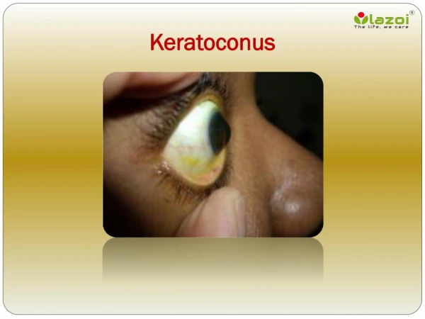
1. Keratoconus
CORNEAL ECTASIAS. 1. Keratoconus. 2. Keratoglobus. 3. Pellucid marginal degeneration. Morphological classification of keratoconus. Nipple cone. Oval cone . Globus cone . Largest. Small and steep curvature. Larger and ellipsoidal. Signs of keratoconus.

1. Keratoconus
E N D
Presentation Transcript
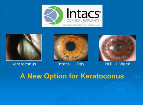
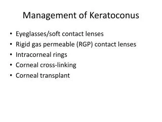
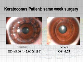
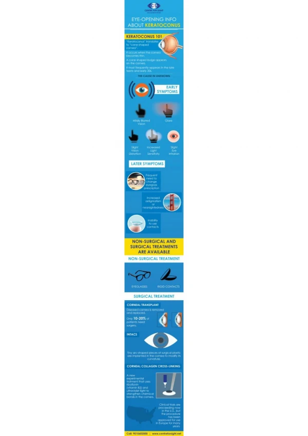
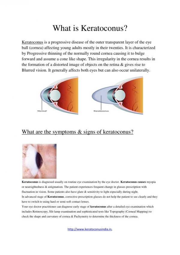
CORNEAL ECTASIAS 1. Keratoconus 2. Keratoglobus 3. Pellucid marginal degeneration
Morphological classification of keratoconus Nipple cone Oval cone Globus cone Largest Small and steep curvature Larger and ellipsoidal
Signs of keratoconus Bilateral in 85% but asymmetrical Oil droplet reflex Vogt striae Prominent corneal nerves Bulging of lower lids on downgaze Munson sign Fleischer ring & scarring Acute hydrops
Systemic associations of keratoconus Atopic dermatitis Down syndrome Ehlers-Danlos syndrome Marfan syndrome Crouzon syndrome Osteogenesis imperfecta
Keratoglobus • Onset usually at birth • Bilateral protrusion and thinning of entire cornea • Associations - Leber congenital amaurosis and blue sclera
Pellucid marginal degeneration • Onset between 20 and 40 years • Bilateral crescent-shaped inferior corneal thinning