Download
1 / 73
770 likes | 1.11k Vues
HEMOPHILIA AND OTHER COAGULOPATHIES. Prof. Dr. Filiz Bakar Yeditepe Üniversitesi Tıp Fakültesi Çocuk Sağlığı ve Hastalıkları Anabilim Dalı. Outline. Normal hemostasis Hemophilia Differential diagnostic tests to coagulation disorders Treatment. Normal hemostasis. HEMOSTATIC MECHANISMS.

E N D
HEMOPHILIA AND OTHER COAGULOPATHIES Prof. Dr. Filiz Bakar Yeditepe Üniversitesi Tıp Fakültesi Çocuk Sağlığı ve Hastalıkları Anabilim Dalı
Outline • Normal hemostasis • Hemophilia • Differential diagnostic tests to coagulation disorders • Treatment
HEMOSTATIC MECHANISMS • Vascular response • Platelet adhesion • Platelet agregation • Clot formation • Clot stabilization • Limitation of clotting to the site of injury by regulatory anticoagulants • Re-establishment of vascular patency through fibrinolysis • Vascular healing
Stages of hemostasis • Primary hemostasis • Von Willebrand factor, platelets, fibrinogen • Secondary hemostasis • Coagulation cascade • Tertiary hemostasis • Cross-linking of fibrin strands • Clot maturation and wound healing
Anticoagulants • All procoagulant proteins are balanced by an anticoagulant protein. • Four clinically important anticoagulants: • Antithrombin III X F Xa and thrombin • Protein C X F Va and F VIIIa • Protein S • Tissue factor pathway inhibitor X F Xa
Definition • Hemophilias are a group of related bleeding disorders that most commonly are inherited • Inherited bleeding disorders include: • Abnormalities of coagulation factors • Abnormalities of platelet function • The term ‘hemophilia’ refers • Factor VIII deficiency (hemophilia A) • Factor IX deficiency (hemophilia B)
Incidince • Combinedincidence of hemophilia A and B is 1/5000 • 80% hemophilia A; 2/3 have severe disease • Hemophilia B; ½ severe disease
Mild : > 5% factor level • Moderate: 1–5% factor level • Severe: < 1% factor level • Within each kindred, similar severity of disease • Multiple genetic defects • Factor IX > 800 • Factor VIII > 700
Genetics • Hemophilia A F VIII (X-linked) • Hemophilia B F IX (X-linked) • Hemophilia C F XI (Autosomal)
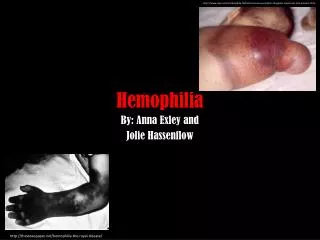
Clinical manifestations • Bleeding may occur anywhere • Most common sites: • Joints (80%) • Ankles; most common in children • Knees, elbows, and ankles; most common in adolescents • Muscles • Gastrointestinal tract
Mild • Bleeding only with response to injury/trauma or surgery • Moderate • Bleeding with response to intercurrent injury or surgery • Severe • Spontaneously and at an early age
Initial presentation • Neonates; • Intracranial hemorrhage • Bleed with circumcision • Infant; • easy bruising, intramuscular hematomas, hemarthroses • Older child; • hemarthroses
Sites of bleeding • Central nervous system • Most frequent in neonates, spontaneous • Hemarthrosis • Painful and physically debilitating manifestation • Originates from the synovial vessels • Hemorrhage occurs within the joint cavity • Hallmark is hemarthroses (ankle, knees, elbows) • In severe hemophilia ‘target joint’
Sites of bleeding • Skeletal muscle • Bleeding into muscles with hematoma formation, most often quadriceps, iliopsoas (important), and forearm • Head and neck • May be life-threatening hemorrhages • Epistaxis, mucosal bleeding, posterior pharynx • Gastrointestinal tract • Genitourinary tract • Hematuria • Posttraumatic bleeding
Late complications • Joint destruction due to hemarthroses • Hemophilic arthropathy; tissue deposition of iron and dense fibrozis of the joint with contractures, pain, limitation of motion • Transmission of blood-borne infections • Hepatitis A, B, C, and D, HIV • Development of inhibitor antibodies • 25% of patients with severe hemophilia A, 3-5% with severe hemophilia B • Complicate bleeding episodes
Hemophilia C • F XI deficiency • Autosomal recessive, frequently in Ashkenazi Jews • Mild-moderate bleeding, not correlated with the amount of F XI.
Deficiencies of the Contact Factors • Extremely prolonged PTT, but no evidence of clinical bleeding • Factor XII, prekallikrein, high molecular weight kininogen
Other Factor Deficiencies • F VII deficiency, AR • F X deficiency, AR • F II (Prothrombin) deficiency, AR • F V deficiency (parahemophilia), AR
Fibrinogen disorders • Severe or mild. • Congenital afibrinogenemia • Rare, AR • Clinical manifestations similar to hemophilia A • Bleeding from minor cuts is prolonged because of the lack of fibrinogen to support platelet aggregation. • Hypofibrinogenemia (AD or AR) • Dysfibrinogenemia (AD) • Mild to moderate bleeding symptoms or may be asymptomatic • Risk for venous thrombosis. • Diagnosis is based on kinetic and antigenic protein levels, thrombin time is usually prolonged
Factor XIII Deficiency • F XIII, responsible for cross linking of fibrin or the stabilization of fibrin clot • Delayed and prolonged bleeding • Autosomal recessive • Clinical symptoms • Umblical bleeding • Intracranial bleeding • Poor wound healing • Delayed separation of umblical cord • Coagulation tests are normal • Clot solubility test (5 M urea)
Von Willebrand disease • Most common hereditary bleeding disorder, incidence 1-2% of general population • Autosomal • Function: Platelet adhesion, F VIII stability • Mucocuteneous hemorrhage, excessive bruising, epistaxis, menorrhagia, postoperative bleeding • Laboratory: long bleeding time, long PTT • Classified as: • Tip 1 (Partial deficiency), 85% • Tip 2 (Functional defect) • Tip 3 (Complete vWF deficiency)
Hereditary predisposition to thrombosis • The newborn infant, because of physiologic deficiency, particulary predisposed to thrombosis. • Deficiencies of regulatory proteins: • Protein C (homozygous protein C deficiency, fatal purpura fulminans in the neonatal period) • Protein S • AT-III • Plasminogen • Synthesis of a procoagulant protein: • F V Leiden • Elevated levels of procoagulant protein: • Prothrombin mutation • Elevated levels of toxic organic acid: • Homocystinuria
Clinical diagnosis • A coagulation disorder is suspected with any of the following: • Hemarthrosis, especially with mild or no antecedent trauma • Deep-muscle hematomas • Intracranial bleeding in the absence of major trauma • Cephalohematoma or intracranial bleeding at birth • Prolonged oozing or renewed bleeding after initial bleeding stops following tooth extractions, mouth injury, or circumcision • Prolonged bleeding or renewed bleeding following surgery or trauma • Unexplained GI bleeding or hematuria • Menorrhagia, especially at menarche • Prolonged nosebleeds, especially recurrent and bilateral • Excessive bruising, especially with firm, subcutaneous hematomas
History • To differentiate hereditary disorders from acquired • Hereditary • Start early in age • Recurrent in nature • Positive family history • Acquired • Occur at any age • Underlying predisposing cause
Physical Examination • Mucocutaneous bleeding (mucous membranes or skin) OR • Deep bleeding (muscles and joints) • Presence of: • petechia, • ecchymoses, • hematomas, • hemartroses, • mucous membrane bleeding
Type of Bleeding for Differential Diagnosis Unilateral epistaxis Non-hematologic Bruises arms and legs Mucose membrane bleeding Platelet disorder, vWD Bleeding into joint and soft tissue Hemophilia A, B, vWD Bilateral epistaxis Excessive menstruel bleeding Umblical bleeding F XIII deficiency
Laboratory Investigation in Patients with Bleeding Diathesis • CBC (platelet count) • Bleeding time • Prothrombin time (PT) • Activated partial thromboplastin time(aPTT) If the results are normal: • Thrombin Time (TT) • vWF testing • Clot stability Further spesific work-up: • F XIII • Factor Activity • Platelet Functions • Antiphospholipid antibodies