Radiology Class Presentation Mammography
Radiology Class Presentation Mammography. Arun Ganguly Ph.D. (Rebecca Fahrig, Ph.D.). Numbers you Need to Know : Dose. The ACR recommends that the AGD should <3mGy (300 mrad ) per film ie . per view for screen/film with a grid (< 1 mGy for no grid)


Radiology Class Presentation Mammography
E N D
Presentation Transcript
Radiology Class Presentation Mammography Arun Ganguly Ph.D. (Rebecca Fahrig, Ph.D.)
Numbers you Need to Know : Dose • The ACR recommends that the AGD should <3mGy (300 mrad) per film ie. per view for screen/film with a grid (< 1 mGy for no grid) • Exposing 1 million 45-y.o. women to 1 mGy causes 2 excess breast cancer deaths • Dose for a typical screening exam is 2.5 mGy, therefore 5 excess deaths per million • Screening : 1 million women results in identification of 3000 cases of breast cancer • Without screening : 50% mortality • Fatality rate is reduced by 40%, or saves 600 lives
Numbers you Need to Know : MQSA (Mammography Quality Standards Act • FDA developed MQSA, passed in 1994, final in 1999. • MUST be certified by the FDA, and therefore accredited by the ACR • Accredited physicians must have interpreted at least 200 mammograms in the last 24 months • ACR requirements: site survey, assess image quality using a phantom, dosimeter of MGD, asses clinical images by independent radiologists, assess quality control program
Things you Need to Know: MQSA • Processor QC on a daily basis • Screens and darkroom cleaned daily • Weekly tests with ACR phantom • Hardware inspected every month • Quarterly tests including repeat analysis (2-5%), fixer retention on film • Darkroom fog, screen/film contact and compression every 6 months
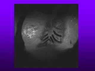
Things you Need to Know : MQSA • Medical physicist tests annually: unit assembly and cassette performance, collimation, system resolution, peak voltage accuracy and reproducibility, beam quality (HVL), AEC performance, uniformity of screen speeds, radiation output, entrance skin exposure and MGD, image quality (phantom), artifact evaluation • ACR phantom : contains fibers (6), speck groups (5) and masses (5) • To PASS : see 4, 3 and 3 • X-ray output must be greater than 7 mGy/s, averaged over 3s • Spatial resolution from focal spot blur must be no worse than 11 lp/mm parallel to anode-cathode axis, 13 lp/mm perpendicular • AEC should maintain film optical density within 0.15 of the mean density
Image Quality Test Objects • SMPTE pattern : digital image with stripes of different contrast used for QC of laser images, monitors etc. • System resolution : lp phantom, MTF measurement • Focal spot size : star pattern, slit system • Film processor : stepped film exposure system, sensitomtric strip • HVL : Al filters
List the following types of x-ray equipment in order of increasing beam HVL? CT, mammo, barium fluoro, skeletal radiography Mammo, skeletal radiography, CT, barium fluoro Barium fluoro, mammo, skeletal radiography, CT Skeletal radiography, bariumfluoro, CT, mammo Mammo, skeletal radiography, barium fluoro, CT
ANSWER: E. Mammo, skeletal radiography, barium fluoro, CT Mammo is done at very low kVp with HVL of 0.3-0.4mm Al, skeletal radiography at 60-70 kVp with HVL of 1.5-2.5mm Al, barium fluoro is at somewhat higher kVp with HVL of 3-5mm Al, whiel CT is performed at 120-140 kVp with added shaped filters giving HVLs of 6mm of Al.
What are the typical focal spot sizes and mA in magnification mammography? 0.1mm 30mA 0.1mm 100mA 0.3mm 30mA 0.3mm 100mA 0.6mm 100mA
ANSWER: A. 0.1mm and 30mA 0.1mm is the smallest focal spot used in radiology and even so it generally does not result in any significant improvement in resolution over the contact 0.3mm focus because of the large 1.8-2.0 fold magnification used in mammography. The highest mA possible for 0.1mm focus is typically 30mA. The mA is reduced to lower the heat generated in such a small size focus.
What artifacts are seen in both full-field digital mammography and film screen mammography? Grid lines Pick-offs Fingerprints Chatter Runback
ANSWER: A. Grid-lines Grids are used in both digital and screen-film mammography. All other s are screen-film artifacts (pick offs: caused due to dirty roller)
In a mammography unit using 100mA, the AEC in Auto kVp mode generally increases the mAs with increasing breast thickness. After about 200mAs, it starts to increase the kVp. Why does the kVp rather than the mAs increase at that point? To maximize contrast for thick breasts To minimize motion blur by not allowing excessively long exposure times To minimize noise To prevent overheating of x-ray tube To reduce the low-energy x-rays which contribute mainly to the dose.
ANSWER: B. To minimize motion blur by not allowing excessively long exposure times The AEC is generally designed to give maximum exposure time of 1.5-2s to minimize motion.
The purpose of the Mo, Rh, or Ag filter in mammography is to filter out? The low-energy x-rays in the useful beam The high-energy x-ray in the useful beam The low and high energy x-rays in the useful beam All but the characteristic x-rays in the useful beam The noise in the image.
ANSWER: C. The low and high energy x-rays in the useful beam All filters remove the low-energy x-rays which do not make it through the breast and only contribute to dose. Mo, Rh, and Ag all have k-edges in the mammographic kVp range (20, 23, 25.5keV), and strongly filter out the high-energy x-rays above the k-edge that would otherwise reduce subject contrast. (for screen film optimal contrast is at 19 keV , thickness dependence)
In full field digital mammography unit, what would result from changing the AEC fine density control from 0 to +1? Dose decreases by 50% Dose decreases by 15% No change in dose, but image brightness increases Dose increases by 15% Dose increases by 50%
ANSWER: D. The dose increases by 15% The ACR Mammography Accreditation QC manual recommends that each step of the density control should change the mAs (and hence the dose) by 12% to 15%
What is the glandular/adipose tissue mix assumed for mammography dosimetry? 100 – 0 75 -25 50-50 25-75 0-100
ANSWER: C. 50-50 This has been the accepted arbitrary convention used for more than 40 years.
What is not an advantage from firm compression in mammography? Lower patient dose Patient discomfort Less scatter Shorter exposure time, less motion blur Less magnification, less geometric blur
ANSWER: B. Patient discomfort All the others (and more) are benefits of firm compression.
Compare typical breast dose from mammo screening 2 views exam and a non-contrast chest CT (per breast)? Mammo=1.5 mGy; CT=1 mGy Mammo=3.0 mGy; CT=1 mGy Mammo=1.5 mGy; CT=10 mGy Mammo=3.0 mGy; CT=10 mGy Mammo=10mGy; CT=10 mGy
ANSWER: D. Mammo=3.0 mGy; CT=10 mGy The average glandular dose in screening mammography for an average 4.5cm compressed breast in the US is 1.5-1.8 mGy per view. A screening exam requires 2 views. A chest CT scan has a typical peripheral dose of about 10 mGy.
What is the advantage of digital tomosynthesis in mammography? Lowest breast dose Thin CT-like slices that remove overlying anatomy Shorter exposure time Better spatial resolution Places marks on suspicious areas in the image
ANSWER: B. Thin CT-like slices that remove overlying anatomy It produces CT-like 1mm slices by taking 15-30 low dose images over 15-30 angular degrees of travel of the x-ray tube. Dose is not increase but resolution may be degraded slightly, mainly because of long exposure time. CAD places marks on suspicious areas in an image.
Which of the following is currently (2008) not regulated by the Mammography Quality Standards Act • CME of reading radiologists • Continuing experience of radiologist • CME of medical physicists • Continuing experience of medical physicists • Average glandular dose in stereotactic breast biopsy
ANSWER: E. Average glandular dose in stereotactic breast biopsy All are regulated by MQSA except stereotactic breast biopsy, which in 2008 is only regulated by the state Departments of Health
Digital mammograms are read on a hospital’s PACS. What is the requirement for the reading station display? • They must be supplied by the digital mammography unit’s manufacturer • They must be flat panel technology • They must be at least 5 megapixels (MPs) • They must be capable of displaying color • There is no requirement; they only need the radiologist’s approval
ANSWER: C. They must be at least 5 megapixels All full-field digital mammography, including CR mammography, must be interpreted on 5 MP displays or laser hardcopy as per MQSA
Digital mammography QC must be performed by technologists and medical physicists. The specific tasks and their frequency of performance are specified by: • The mammography equipment manufacturer • The ACR Mammography Accreditation Program • MQSA • The state Departments of Health • The medical physicist
ANSWER: A. The equipment manufacturer for full-field digital mammography, MQSA and ACR require the QC tasks and frequencies to be those specified by the FFDM manufacturer
Digital tomosynthesis is used in mammography to: • Improve spatial resolution • Improve contrast in lesions • Reduce motion blurring • Reduce radiation dose • Eliminate the need for compression
ANSWER: B. Improve contrast in lesions Tomosynthesis provides thin slices that reduce superpositioning of structures (ie., volume averaging) and thus improves contrast of small lesions. Resolution may decrease because of imperfect motion of the tube-receptor. Motion blurring in the patient may increase because of the long scan time. Radiation dose will probably increase because of the need to reduce noise in the thin slices.
A major limitation of many early full-field digital mammography units is the ____: • Focal spot size • Detector size • Resolution capability • Lack of compression capability • Limited angulation
ANSWER: B. Detector size. In the early FFDM units the detector size was typically about 18x24cm, similar to the smaller size mammography film cassettes. When patients with large breast were imaged, double exposures were often necessary in order to fully image the large breast. The two images then had to be viewed sequentially in order to get information on the one breast. Some newer units have larger detectors, capable of imaging approximately 24x30 cm, similar to the larger size mammography film cassettes. The compression & angulation capabilities are essentially the same as on film units. The possible lower resolution of the digital units does not appear to significantly affect diagnosis.
In mammography, what would an SMPTE pattern be used for? • Viewbox QC • Evaluation of system resolution • Evaluation of focal spot size • QC of laser image printer & display monitor • Film processor QC
ANSWER: D. QC of laser image printer & display monitor The SMPTE pattern is a standard digital image with steps of different contrast used for QC & setup of laser imagers (digital mammography) and image viewing monitors.
If all of the following mammography techniques result in the same OD (~1.8) for a certain breast thickness, which is the preferred technique? • 24 kVp, 5 s • 26 kVp, 3 s • 28 kVp, 1.5 s • 31 kVp, 0.8 s • 34 kVp, 0.4 s
ANSWER: C. 28 kVp, 1.5 s The lowest kVp (for good contrast) that results in an exposure time between 1 & 2 seconds (for minimum motion blur) should be used. Exposure times in A & B are too long (increased blur). In D & E, the kVp is higher than necessary (reduced contrast).
MQSA-required mammography screen-film system resolution is 11 lp/mm. The typical system resolution for FFDM (Full Field Digital Mammography) is ____lp/mm. • 25 • 20 • 15 • 11 • 5
ANSWER: E. 5. FFDM has a considerably lower system resolution than screen-film because of the rather large pixel size of the digital detectors (0.05-0.1 mm). However, FFDM sensitivity to detect cancer has been shown to be at least equivalent to screen-film systems.
Which of the following is nottrue about the average glandular dose (AGD) for sterotactic breast biopsy (SBB)? • MQSA limits the SBB AGD to 3mGy for an average breast. • The ACR SBB accreditation program recommends <3 mGy for an average breast. • Some states limit the SBB AGD to 3 mGy for an average breast. • The SBB AGD is calculated in the same way as for whole breast mammography. • There is no limit on the SBB AGD for a thick, dense breast.
ANSWER: A. MQSA limits the SBB AGD to 3mGy for an average breast. MQSA (as of 2005) does not regulate SBB at all. Some states do regulate SBB & generally follow the recommendations of the ACR SBB accreditation program, which recommends an AGD of less than 3 mGy for an average breast.
Breast microcalcifications are seldom seen on routine chest radiographs because: • The film size is too large. • Of an increased mass attenuation coefficient for soft tissue. • Of a decreased mass attenuation coefficient for soft tissue. • Of an increased mass attenuation coefficient for calcium. • Of an decreased mass attenuation coefficient for calcium.